- Bioactive Compounds
- By Signaling Pathways
- PI3K/Akt/mTOR
- Epigenetics
- Methylation
- Immunology & Inflammation
- Protein Tyrosine Kinase
- Angiogenesis
- Apoptosis
- Autophagy
- ER stress & UPR
- JAK/STAT
- MAPK
- Cytoskeletal Signaling
- Cell Cycle
- TGF-beta/Smad
- Compound Libraries
- Popular Compound Libraries
- Customize Library
- Clinical and FDA-approved Related
- Bioactive Compound Libraries
- Inhibitor Related
- Natural Product Related
- Metabolism Related
- Cell Death Related
- By Signaling Pathway
- By Disease
- Anti-infection and Antiviral Related
- Neuronal and Immunology Related
- Fragment and Covalent Related
- FDA-approved Drug Library
- FDA-approved & Passed Phase I Drug Library
- Preclinical/Clinical Compound Library
- Bioactive Compound Library-I
- Bioactive Compound Library-Ⅱ
- Kinase Inhibitor Library
- Express-Pick Library
- Natural Product Library
- Human Endogenous Metabolite Compound Library
- Alkaloid Compound LibraryNew
- Angiogenesis Related compound Library
- Anti-Aging Compound Library
- Anti-alzheimer Disease Compound Library
- Antibiotics compound Library
- Anti-cancer Compound Library
- Anti-cancer Compound Library-Ⅱ
- Anti-cancer Metabolism Compound Library
- Anti-Cardiovascular Disease Compound Library
- Anti-diabetic Compound Library
- Anti-infection Compound Library
- Antioxidant Compound Library
- Anti-parasitic Compound Library
- Antiviral Compound Library
- Apoptosis Compound Library
- Autophagy Compound Library
- Calcium Channel Blocker LibraryNew
- Cambridge Cancer Compound Library
- Carbohydrate Metabolism Compound LibraryNew
- Cell Cycle compound library
- CNS-Penetrant Compound Library
- Covalent Inhibitor Library
- Cytokine Inhibitor LibraryNew
- Cytoskeletal Signaling Pathway Compound Library
- DNA Damage/DNA Repair compound Library
- Drug-like Compound Library
- Endoplasmic Reticulum Stress Compound Library
- Epigenetics Compound Library
- Exosome Secretion Related Compound LibraryNew
- FDA-approved Anticancer Drug LibraryNew
- Ferroptosis Compound Library
- Flavonoid Compound Library
- Fragment Library
- Glutamine Metabolism Compound Library
- Glycolysis Compound Library
- GPCR Compound Library
- Gut Microbial Metabolite Library
- HIF-1 Signaling Pathway Compound Library
- Highly Selective Inhibitor Library
- Histone modification compound library
- HTS Library for Drug Discovery
- Human Hormone Related Compound LibraryNew
- Human Transcription Factor Compound LibraryNew
- Immunology/Inflammation Compound Library
- Inhibitor Library
- Ion Channel Ligand Library
- JAK/STAT compound library
- Lipid Metabolism Compound LibraryNew
- Macrocyclic Compound Library
- MAPK Inhibitor Library
- Medicine Food Homology Compound Library
- Metabolism Compound Library
- Methylation Compound Library
- Mouse Metabolite Compound LibraryNew
- Natural Organic Compound Library
- Neuronal Signaling Compound Library
- NF-κB Signaling Compound Library
- Nucleoside Analogue Library
- Obesity Compound Library
- Oxidative Stress Compound LibraryNew
- Plant Extract Library
- Phenotypic Screening Library
- PI3K/Akt Inhibitor Library
- Protease Inhibitor Library
- Protein-protein Interaction Inhibitor Library
- Pyroptosis Compound Library
- Small Molecule Immuno-Oncology Compound Library
- Mitochondria-Targeted Compound LibraryNew
- Stem Cell Differentiation Compound LibraryNew
- Stem Cell Signaling Compound Library
- Natural Phenol Compound LibraryNew
- Natural Terpenoid Compound LibraryNew
- TGF-beta/Smad compound library
- Traditional Chinese Medicine Library
- Tyrosine Kinase Inhibitor Library
- Ubiquitination Compound Library
-
Cherry Picking
You can personalize your library with chemicals from within Selleck's inventory. Build the right library for your research endeavors by choosing from compounds in all of our available libraries.
Please contact us at info@selleckchem.com to customize your library.
You could select:
- Antibodies
- Bioreagents
- qPCR
- 2x SYBR Green qPCR Master Mix
- 2x SYBR Green qPCR Master Mix(Low ROX)
- 2x SYBR Green qPCR Master Mix(High ROX)
- Protein Assay
- Protein A/G Magnetic Beads for IP
- Anti-DYKDDDDK Tag magnetic beads
- Anti-DYKDDDDK Tag Affinity Gel
- Anti-Myc magnetic beads
- Anti-HA magnetic beads
- Poly DYKDDDDK Tag Peptide lyophilized powder
- Protease Inhibitor Cocktail
- Protease Inhibitor Cocktail (EDTA-Free, 100X in DMSO)
- Phosphatase Inhibitor Cocktail (2 Tubes, 100X)
- Cell Biology
- Cell Counting Kit-8 (CCK-8)
- Animal Experiment
- Mouse Direct PCR Kit (For Genotyping)
- New Products
- Contact Us
Quisinostat (JNJ-26481585) 2HCl
Quisinostat (JNJ-26481585) 2HCl is a novel second-generation HDAC inhibitor with highest potency for HDAC1 with IC50 of 0.11 nM in a cell-free assay, modest potent to HDACs 2, 4, 10, and 11; greater than 30-fold selectivity against HDACs 3, 5, 8, and 9 and lowest potency to HDACs 6 and 7. Phase 2.

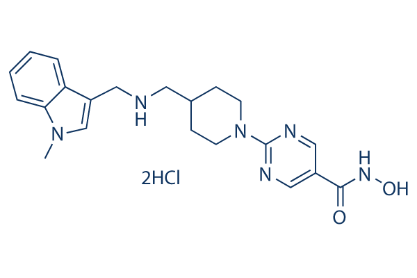
Quisinostat (JNJ-26481585) 2HCl Chemical Structure
CAS: 875320-31-3
Selleck's Quisinostat (JNJ-26481585) 2HCl has been cited by 57 publications
Purity & Quality Control
Batch:
Purity:
99.62%
99.62
Other HDAC Products
Related compound libraries
Choose Selective HDAC Inhibitors
Cell Data
| Cell Lines | Assay Type | Concentration | Incubation Time | Formulation | Activity Description | PMID |
|---|---|---|---|---|---|---|
| A549 cells | Cell viability assay | 24, 48, or 72 h | the IC50 values of cells for 48 and 72 h of quisinostat treatment were 82.4 and 42.0 nM, respectively. | 27423454 | ||
| RH30 cells | Function assay | 15 nM | 24 h and 30 h | GSK690 and JNJ-26481585 cooperated to induce cleavage of the initiator caspase-9 into its active p35 and p37 fragments and of the effector caspase-3 into its active p12 and p17 fragments | 28617441 | |
| RD cells | Function assay | 15 nM | 24 h and 30 h | GSK690 and JNJ-26481585 cooperated to induce cleavage of the initiator caspase-9 into its active p35 and p37 fragments and of the effector caspase-3 into its active p12 and p17 fragments | 28617441 | |
| HepG2 | Cell viability assay | 5, 10, 20, 40, 80, 160 and 320 nM | 24, 48, 72 h | The IC50 values of cells for quisinostat treatment at 48 and 72 h were 81.2 and 30.8 nM, respectively. | 28849080 | |
| HuT78 | Function assay | 10 nM | increases the level of acetylated lysine K10 of histone H3 | 30012418 | ||
| Sf9 | Function assay | Inhibition of full length recombinant human HDAC1 expressed in baculovirus infected Sf9 cells using RHKK-Ac as substrate by fluorescence analysis, IC50 = 0.00011 μM. | 27606546 | |||
| Sf9 | Function assay | Inhibition of full length recombinant human HDAC2 expressed in baculovirus infected Sf9 cells using RHKK-Ac as substrate by fluorescence analysis, IC50 = 0.00033 μM. | 27606546 | |||
| Sf9 | Function assay | Inhibition of full length recombinant human HDAC3/NCOR2 expressed in baculovirus infected Sf9 cells using RHKK-Ac as substrate by fluorescence analysis, IC50 = 0.00486 μM. | 27606546 | |||
| HUT78 | Function assay | 18 hrs | Pro-apoptotic activity in human HUT78 cells after 18 hrs by caspase-Glo 3/7 assay, EC50 = 0.0065 μM. | 30122227 | ||
| BL21 (DE3) | Function assay | Binding affinity to human His-thioredoxin-tagged HDAC8 expressed in Escherichia coli BL21 (DE3) cells by ITC method, Kd = 0.0227 μM. | 30347148 | |||
| BL21 (DE3) | Function assay | Binding affinity to Schistosoma mansoni His-tagged HDAC8 expressed in Escherichia coli BL21 (DE3) cells by ITC method, Kd = 0.0284 μM. | 30347148 | |||
| BL21 (DE3) | Function assay | Inhibition of human His-thioredoxin-tagged HDAC8 expressed in Escherichia coli BL21 (DE3) cells using Fluor de Lys (R)-HDAC8 as substrate by fluorometric method, IC50 = 0.0644 μM. | 30347148 | |||
| Sf9 | Function assay | Inhibition of full length recombinant human HDAC6 expressed in baculovirus infected Sf9 cells using RHKK-Ac as substrate by fluorescence analysis, IC50 = 0.0768 μM. | 27606546 | |||
| BL21 (DE3) | Function assay | Inhibition of human His-thioredoxin-tagged HDAC8 mL6 mutant expressed in Escherichia coli BL21 (DE3) cells using Fluor de Lys (R)-HDAC8 as substrate by fluorometric method, IC50 = 0.093 μM. | 30347148 | |||
| BL21 (DE3) | Function assay | Inhibition of human His-thioredoxin-tagged HDAC8 mL1/mL6 mutant expressed in Escherichia coli BL21 (DE3) cells using Fluor de Lys (R)-HDAC8 as substrate by fluorometric method, IC50 = 0.094 μM. | 30347148 | |||
| BL21 (DE3) | Function assay | Inhibition of human His-thioredoxin-tagged HDAC8 mL6/L179I mutant expressed in Escherichia coli BL21 (DE3) cells using Fluor de Lys (R)-HDAC8 as substrate by fluorometric method, IC50 = 0.169 μM. | 30347148 | |||
| BL21 (DE3) | Function assay | Inhibition of human His-thioredoxin-tagged HDAC8 mL1/mL6/L179I mutant expressed in Escherichia coli BL21 (DE3) cells using Fluor de Lys (R)-HDAC8 as substrate by fluorometric method, IC50 = 0.217 μM. | 30347148 | |||
| BL21 (DE3) | Function assay | Inhibition of Schistosoma mansoni His-tagged HDAC8 expressed in Escherichia coli BL21 (DE3) cells using Fluor de Lys (R)-HDAC8 as substrate by fluorometric method, IC50 = 0.3034 μM. | 30347148 | |||
| TC32 | qHTS assay | qHTS of pediatric cancer cell lines to identify multiple opportunities for drug repurposing: Primary screen for TC32 cells | 29435139 | |||
| U-2 OS | qHTS assay | qHTS of pediatric cancer cell lines to identify multiple opportunities for drug repurposing: Primary screen for U-2 OS cells | 29435139 | |||
| A673 | qHTS assay | qHTS of pediatric cancer cell lines to identify multiple opportunities for drug repurposing: Primary screen for A673 cells | 29435139 | |||
| DAOY | qHTS assay | qHTS of pediatric cancer cell lines to identify multiple opportunities for drug repurposing: Primary screen for DAOY cells | 29435139 | |||
| Saos-2 | qHTS assay | qHTS of pediatric cancer cell lines to identify multiple opportunities for drug repurposing: Primary screen for Saos-2 cells | 29435139 | |||
| BT-37 | qHTS assay | qHTS of pediatric cancer cell lines to identify multiple opportunities for drug repurposing: Primary screen for BT-37 cells | 29435139 | |||
| RD | qHTS assay | qHTS of pediatric cancer cell lines to identify multiple opportunities for drug repurposing: Primary screen for RD cells | 29435139 | |||
| SK-N-SH | qHTS assay | qHTS of pediatric cancer cell lines to identify multiple opportunities for drug repurposing: Primary screen for SK-N-SH cells | 29435139 | |||
| BT-12 | qHTS assay | qHTS of pediatric cancer cell lines to identify multiple opportunities for drug repurposing: Primary screen for BT-12 cells | 29435139 | |||
| MG 63 (6-TG R) | qHTS assay | qHTS of pediatric cancer cell lines to identify multiple opportunities for drug repurposing: Primary screen for MG 63 (6-TG R) cells | 29435139 | |||
| NB1643 | qHTS assay | qHTS of pediatric cancer cell lines to identify multiple opportunities for drug repurposing: Primary screen for NB1643 cells | 29435139 | |||
| OHS-50 | qHTS assay | qHTS of pediatric cancer cell lines to identify multiple opportunities for drug repurposing: Primary screen for OHS-50 cells | 29435139 | |||
| BT-12 | qHTS assay | qHTS of pediatric cancer cell lines to identify multiple opportunities for drug repurposing: Confirmatory screen for BT-12 cells | 29435139 | |||
| DAOY | qHTS assay | qHTS of pediatric cancer cell lines to identify multiple opportunities for drug repurposing: Confirmatory screen for DAOY cells | 29435139 | |||
| fibroblast cells | qHTS assay | qHTS of pediatric cancer cell lines to identify multiple opportunities for drug repurposing: Primary screen for control Hh wild type fibroblast cells | 29435139 | |||
| SK-N-SH | qHTS assay | qHTS of pediatric cancer cell lines to identify multiple opportunities for drug repurposing: Confirmatory screen for SK-N-SH cells | 29435139 | |||
| Rh41 | qHTS assay | qHTS of pediatric cancer cell lines to identify multiple opportunities for drug repurposing: Primary screen for Rh41 cells | 29435139 | |||
| A673 | qHTS assay | qHTS of pediatric cancer cell lines to identify multiple opportunities for drug repurposing: Confirmatory screen for A673 cells) | 29435139 | |||
| Rh30 | qHTS assay | qHTS of pediatric cancer cell lines to identify multiple opportunities for drug repurposing: Primary screen for Rh30 cells | 29435139 | |||
| MG 63 (6-TG R) | qHTS assay | qHTS of pediatric cancer cell lines to identify multiple opportunities for drug repurposing: Confirmatory screen for MG 63 (6-TG R) cells | 29435139 | |||
| fibroblast cells | qHTS assay | qHTS of pediatric cancer cell lines to identify multiple opportunities for drug repurposing: Confirmatory screen for control Hh wild type fibroblast cells | 29435139 | |||
| Rh30 | qHTS assay | qHTS of pediatric cancer cell lines to identify multiple opportunities for drug repurposing: Confirmatory screen for Rh30 cells | 29435139 | |||
| U-2 OS | qHTS assay | qHTS of pediatric cancer cell lines to identify multiple opportunities for drug repurposing: Confirmatory screen for U-2 OS cells | 29435139 | |||
| OHS-50 | qHTS assay | qHTS of pediatric cancer cell lines to identify multiple opportunities for drug repurposing: Confirmatory screen for OHS-50 cells | 29435139 | |||
| SK-N-SH | qHTS assay | qHTS of pediatric cancer cell lines to identify multiple opportunities for drug repurposing: Orthogonal 3D viability screen for SK-N-SH cells | 29435139 | |||
| Rh41 | qHTS assay | qHTS of pediatric cancer cell lines to identify multiple opportunities for drug repurposing: Confirmatory screen for Rh41 cells | 29435139 | |||
| RD | qHTS assay | qHTS of pediatric cancer cell lines to identify multiple opportunities for drug repurposing: Confirmatory screen for RD cells | 29435139 | |||
| Daoy | qHTS assay | qHTS of pediatric cancer cell lines to identify multiple opportunities for drug repurposing: Orthogonal 3D viability screen for Daoy cells | 29435139 | |||
| TC32 | qHTS assay | qHTS of pediatric cancer cell lines to identify multiple opportunities for drug repurposing: Orthogonal 3D viability screen for TC32 cells | 29435139 | |||
| MG 63 (6-TG R) | qHTS assay | qHTS of pediatric cancer cell lines to identify multiple opportunities for drug repurposing: Orthogonal 3D viability screen for MG 63 (6-TG R) cells | 29435139 | |||
| SJ-GBM2 | qHTS assay | qHTS of pediatric cancer cell lines to identify multiple opportunities for drug repurposing: Primary screen for SJ-GBM2 cells | 29435139 | |||
| SK-N-MC | qHTS assay | qHTS of pediatric cancer cell lines to identify multiple opportunities for drug repurposing: Primary screen for SK-N-MC cells | 29435139 | |||
| NB-EBc1 | qHTS assay | qHTS of pediatric cancer cell lines to identify multiple opportunities for drug repurposing: Primary screen for NB-EBc1 cells | 29435139 | |||
| LAN-5 | qHTS assay | qHTS of pediatric cancer cell lines to identify multiple opportunities for drug repurposing: Primary screen for LAN-5 cells | 29435139 | |||
| Rh18 | qHTS assay | qHTS of pediatric cancer cell lines to identify multiple opportunities for drug repurposing: Primary screen for Rh18 cells | 29435139 | |||
| NB1643 | qHTS assay | qHTS of pediatric cancer cell lines to identify multiple opportunities for drug repurposing: Confirmatory screen for NB1643 cells | 29435139 | |||
| SJ-GBM2 | qHTS assay | qHTS of pediatric cancer cell lines to identify multiple opportunities for drug repurposing: Confirmatory screen for SJ-GBM2 cells | 29435139 | |||
| Rh18 | qHTS assay | qHTS of pediatric cancer cell lines to identify multiple opportunities for drug repurposing: Confirmatory screen for Rh18 cells | 29435139 | |||
| Saos-2 | qHTS assay | qHTS of pediatric cancer cell lines to identify multiple opportunities for drug repurposing: Confirmatory screen for Saos-2 cells | 29435139 | |||
| SJ-GBM2 | qHTS assay | qHTS of pediatric cancer cell lines to identify multiple opportunities for drug repurposing: Orthogonal 3D viability screen for SJ-GBM2 cells | 29435139 | |||
| RD | qHTS assay | qHTS of pediatric cancer cell lines to identify multiple opportunities for drug repurposing: Orthogonal 3D viability screen for RD cells | 29435139 | |||
| Click to View More Cell Line Experimental Data | ||||||
Biological Activity
| Description | Quisinostat (JNJ-26481585) 2HCl is a novel second-generation HDAC inhibitor with highest potency for HDAC1 with IC50 of 0.11 nM in a cell-free assay, modest potent to HDACs 2, 4, 10, and 11; greater than 30-fold selectivity against HDACs 3, 5, 8, and 9 and lowest potency to HDACs 6 and 7. Phase 2. | |||||||||||
|---|---|---|---|---|---|---|---|---|---|---|---|---|
| Features | An orally bioavailable, second-generation, hydroxamic acid-based HDAC inhibitor. | |||||||||||
| Targets |
|
| In vitro | ||||
| In vitro | JNJ-26481585 exhibits broad spectrum antiproliferative activity in solid and hematologic cancer cell lines, such as all lung, breast, colon, prostate, brain, and ovarian tumor cell lines, with IC50 ranging from 3.1-246 nM, which is more potent than vorinostat, R306465, panobinostat, CRA-24781, or mocetinostat in various human cancer cell lines tested. [1] A recent study shows that JNJ-26481585 promotes myeloma cell death at low nanomolar concentrations by resulting in Mcl-1 depletion and Hsp72 induction. [2] | |||
|---|---|---|---|---|
| Kinase Assay | HDAC activity assays | |||
| In all cases, full-length HDAC proteins are expressed using baculovirus-infected Sf9 cells. In addition, HDAC3 is coexpressed as a complex with human NCOR2. For assessing activity of HDAC1-containing cellular complexes, immunoprecipitated HDAC1 complexes are incubated with an [3H]acetyl- labeled fragment of histone H4 peptide [biotin-(6-aminohexanoic)Gly-Ala-(acetyl[3H])Lys-Arg-His-Arg-Lys-Val-NH2] in a total volume of 50μL enzyme assay buffer (25mM HEPES (pH 7.4), 1 M sucrose, 0.1 mg/mL BSA and 0.01% (v/v) Triton X-100). Incubation is performed for 45 minutes at 37 °C (immunoprecipitates) or 30 min at room temperature. Before addition of substrate, HDAC inhibitors are added at increasing concentrations and preincubated for 10 minutes at room temperature. After incubation, the reaction is quenched with 35μL stop buffer (1 M HCl and 0.4 M acetic acid). Released [3H]acetic acid is extracted with 800μL ethyl acetate and quantified by scintillation counting. Equal amounts of HDAC1 are immunoprecipitated as indicated by Western blot analysis. HDAC1 activity results are presented as mean ± SD of three independent experiments on a single lysate. | ||||
| Cell Research | Cell lines | NCL-H2106, Colo699 and LNCAP cells | ||
| Concentrations | 0-300 nM | |||
| Incubation Time | 24 hours | |||
| Method | All cell lines are obtained from American Type Culture Collection and cultured according to instructions. The effect of HDAC inhibitors on cell proliferation is measured using an MTT. Proliferation of non–small cell lung carcinoma (NSCLC) cell lines is assessed using an Alamar Blue–based assay. For proliferation of hematologic cell lines, cells are incubated for 72 hours and the cytotoxic activity is evaluated by MTS assay. Data are presented as mean IC50 or IC40 ± SD of at least three independent experiments. | |||
| Experimental Result Images | Methods | Biomarkers | Images | PMID |
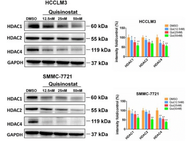
| Western blot | HDAC1 / HDAC2 / HDAC4 p21 / CDK2 / CDK4 / CDK6 / Cyclin D1 / Cyclin E1 / Cyclin A2 Caspase-3/ Cleaved caspase-3 / caspase-9 / Cleaved caspase-9 / PARP / Cleaved PARP / Bcl-xl / Bcl2 / Bax / Survivin PI3K-p110 / PI3K-p85 / p-AKT / JNK / p-JNK / p-c-Jun |

|
30443188 | |
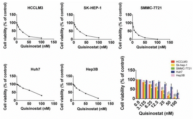
| Growth inhibition assay | Cell viability |

|
30443188 | |
| In Vivo | ||
| In vivo | In an HDAC1-responsive A2780 ovarian tumor screening model, JNJ-26481585 dosing at its maximal tolerated dose (10 mg/kg i.p. and 40 mg/kg p.o.) for 3 days leads to an HDAC1-regulated fluorescence , which predicts tumor growth inhibition. Furthermore, JNJ-26481585 also shows more potent inhibitory effects on the growth of C170HM2 colorectal liver metastases than 5-fluorouracil/Leucovorin. [1] | |
|---|---|---|
| Animal Research | Animal Models | HCT116 human colon carcinoma cells are injected s.c. into the inguinal region of athymic male NMRI nu/nu mice, C170HM2 cell suspensions are injected into the peritoneal cavity of male MFI nude mice. |
| Dosages | ≤10 mg/kg | |
| Administration | Administered via both p.o. and i.p. | |
| NCT Number | Recruitment | Conditions | Sponsor/Collaborators | Start Date | Phases |
|---|---|---|---|---|---|
| NCT02948075 | Completed | Ovarian Cancer | NewVac LLC|Janssen Pharmaceutica N.V. Belgium | September 2015 | Phase 2 |
| NCT01464112 | Completed | Multiple Myeloma | Janssen Research & Development LLC | September 16 2011 | Phase 1 |
| NCT00676728 | Terminated | Advanced or Refractory Leukemia|Myelodysplastic Syndromes | Johnson & Johnson Pharmaceutical Research & Development L.L.C. | December 2008 | Phase 1 |
| NCT00677105 | Completed | Lymphoma|Neoplasms | Johnson & Johnson Pharmaceutical Research & Development L.L.C.|Janssen Pharmaceutica N.V. Belgium | August 2007 | Phase 1 |
Chemical lnformation & Solubility
| Molecular Weight | 467.39 | Formula | C21H28Cl2N6O2 |
| CAS No. | 875320-31-3 | SDF | -- |
| Smiles | CN1C=C(C2=CC=CC=C21)CNCC3CCN(CC3)C4=NC=C(C=N4)C(=O)NO.Cl.Cl | ||
| Storage (From the date of receipt) | |||
|
In vitro |
DMSO : 79 mg/mL ( (169.02 mM); Moisture-absorbing DMSO reduces solubility. Please use fresh DMSO.) Water : Insoluble Ethanol : Insoluble |
Molecular Weight Calculator |
|
In vivo Add solvents to the product individually and in order. |
In vivo Formulation Calculator |
||||
Preparing Stock Solutions
Molarity Calculator
In vivo Formulation Calculator (Clear solution)
Step 1: Enter information below (Recommended: An additional animal making an allowance for loss during the experiment)
mg/kg
g
μL
Step 2: Enter the in vivo formulation (This is only the calculator, not formulation. Please contact us first if there is no in vivo formulation at the solubility Section.)
% DMSO
%
% Tween 80
% ddH2O
%DMSO
%
Calculation results:
Working concentration: mg/ml;
Method for preparing DMSO master liquid: mg drug pre-dissolved in μL DMSO ( Master liquid concentration mg/mL, Please contact us first if the concentration exceeds the DMSO solubility of the batch of drug. )
Method for preparing in vivo formulation: Take μL DMSO master liquid, next addμL PEG300, mix and clarify, next addμL Tween 80, mix and clarify, next add μL ddH2O, mix and clarify.
Method for preparing in vivo formulation: Take μL DMSO master liquid, next add μL Corn oil, mix and clarify.
Note: 1. Please make sure the liquid is clear before adding the next solvent.
2. Be sure to add the solvent(s) in order. You must ensure that the solution obtained, in the previous addition, is a clear solution before proceeding to add the next solvent. Physical methods such
as vortex, ultrasound or hot water bath can be used to aid dissolving.
Tech Support
Answers to questions you may have can be found in the inhibitor handling instructions. Topics include how to prepare stock solutions, how to store inhibitors, and issues that need special attention for cell-based assays and animal experiments.
Tel: +1-832-582-8158 Ext:3
If you have any other enquiries, please leave a message.
* Indicates a Required Field
Frequently Asked Questions
Question 1:
I was thinking of resuspending the powder in 10% hydroxy-propyl-β-cyclodextrin, 25mg/ml mannitol, in sterile water (final pH 8.7). Is this a good vehicle to use? What is the solubility of this chemical in such a vehicle?
Answer:
This vehicle can be used for in vivo studies. The following papers also used this vehicle: 1. http://www.nature.com/leu/journal/v23/n10/full/leu2009121a.html; 2. http://cancerres.aacrjournals.org/content/69/13/5307.long (The solvent contains 10% hydroxypropyl-β-cyclodextrin, 0.8% HCl (0.1 N), 0.9 % NaOH (0.1 N), 3.4% mannitol and pyrogen-free water). The solubility is 2mg/ml.
Tags: buy Quisinostat|Quisinostat ic50|Quisinostat price|Quisinostat cost|Quisinostat solubility dmso|Quisinostat purchase|Quisinostat manufacturer|Quisinostat research buy|Quisinostat order|Quisinostat mouse|Quisinostat chemical structure|Quisinostat mw|Quisinostat molecular weight|Quisinostat datasheet|Quisinostat supplier|Quisinostat in vitro|Quisinostat cell line|Quisinostat concentration|Quisinostat nmr|Quisinostat in vivo|Quisinostat clinical trial|Quisinostat inhibitor|Quisinostat Epigenetics inhibitor


