- Bioactive Compounds
- By Signaling Pathways
- PI3K/Akt/mTOR
- Epigenetics
- Methylation
- Immunology & Inflammation
- Protein Tyrosine Kinase
- Angiogenesis
- Apoptosis
- Autophagy
- ER stress & UPR
- JAK/STAT
- MAPK
- Cytoskeletal Signaling
- Cell Cycle
- TGF-beta/Smad
- Compound Libraries
- Popular Compound Libraries
- Customize Library
- Clinical and FDA-approved Related
- Bioactive Compound Libraries
- Inhibitor Related
- Natural Product Related
- Metabolism Related
- Cell Death Related
- By Signaling Pathway
- By Disease
- Anti-infection and Antiviral Related
- Neuronal and Immunology Related
- Fragment and Covalent Related
- FDA-approved Drug Library
- FDA-approved & Passed Phase I Drug Library
- Preclinical/Clinical Compound Library
- Bioactive Compound Library-I
- Bioactive Compound Library-Ⅱ
- Kinase Inhibitor Library
- Express-Pick Library
- Natural Product Library
- Human Endogenous Metabolite Compound Library
- Alkaloid Compound LibraryNew
- Angiogenesis Related compound Library
- Anti-Aging Compound Library
- Anti-alzheimer Disease Compound Library
- Antibiotics compound Library
- Anti-cancer Compound Library
- Anti-cancer Compound Library-Ⅱ
- Anti-cancer Metabolism Compound Library
- Anti-Cardiovascular Disease Compound Library
- Anti-diabetic Compound Library
- Anti-infection Compound Library
- Antioxidant Compound Library
- Anti-parasitic Compound Library
- Antiviral Compound Library
- Apoptosis Compound Library
- Autophagy Compound Library
- Calcium Channel Blocker LibraryNew
- Cambridge Cancer Compound Library
- Carbohydrate Metabolism Compound LibraryNew
- Cell Cycle compound library
- CNS-Penetrant Compound Library
- Covalent Inhibitor Library
- Cytokine Inhibitor LibraryNew
- Cytoskeletal Signaling Pathway Compound Library
- DNA Damage/DNA Repair compound Library
- Drug-like Compound Library
- Endoplasmic Reticulum Stress Compound Library
- Epigenetics Compound Library
- Exosome Secretion Related Compound LibraryNew
- FDA-approved Anticancer Drug LibraryNew
- Ferroptosis Compound Library
- Flavonoid Compound Library
- Fragment Library
- Glutamine Metabolism Compound Library
- Glycolysis Compound Library
- GPCR Compound Library
- Gut Microbial Metabolite Library
- HIF-1 Signaling Pathway Compound Library
- Highly Selective Inhibitor Library
- Histone modification compound library
- HTS Library for Drug Discovery
- Human Hormone Related Compound LibraryNew
- Human Transcription Factor Compound LibraryNew
- Immunology/Inflammation Compound Library
- Inhibitor Library
- Ion Channel Ligand Library
- JAK/STAT compound library
- Lipid Metabolism Compound LibraryNew
- Macrocyclic Compound Library
- MAPK Inhibitor Library
- Medicine Food Homology Compound Library
- Metabolism Compound Library
- Methylation Compound Library
- Mouse Metabolite Compound LibraryNew
- Natural Organic Compound Library
- Neuronal Signaling Compound Library
- NF-κB Signaling Compound Library
- Nucleoside Analogue Library
- Obesity Compound Library
- Oxidative Stress Compound LibraryNew
- Plant Extract Library
- Phenotypic Screening Library
- PI3K/Akt Inhibitor Library
- Protease Inhibitor Library
- Protein-protein Interaction Inhibitor Library
- Pyroptosis Compound Library
- Small Molecule Immuno-Oncology Compound Library
- Mitochondria-Targeted Compound LibraryNew
- Stem Cell Differentiation Compound LibraryNew
- Stem Cell Signaling Compound Library
- Natural Phenol Compound LibraryNew
- Natural Terpenoid Compound LibraryNew
- TGF-beta/Smad compound library
- Traditional Chinese Medicine Library
- Tyrosine Kinase Inhibitor Library
- Ubiquitination Compound Library
-
Cherry Picking
You can personalize your library with chemicals from within Selleck's inventory. Build the right library for your research endeavors by choosing from compounds in all of our available libraries.
Please contact us at [email protected] to customize your library.
You could select:
- Antibodies
- Bioreagents
- qPCR
- 2x SYBR Green qPCR Master Mix
- 2x SYBR Green qPCR Master Mix(Low ROX)
- 2x SYBR Green qPCR Master Mix(High ROX)
- Protein Assay
- Protein A/G Magnetic Beads for IP
- Anti-DYKDDDDK Tag magnetic beads
- Anti-DYKDDDDK Tag Affinity Gel
- Anti-Myc magnetic beads
- Anti-HA magnetic beads
- Poly DYKDDDDK Tag Peptide lyophilized powder
- Protease Inhibitor Cocktail
- Protease Inhibitor Cocktail (EDTA-Free, 100X in DMSO)
- Phosphatase Inhibitor Cocktail (2 Tubes, 100X)
- Cell Biology
- Cell Counting Kit-8 (CCK-8)
- Animal Experiment
- Mouse Direct PCR Kit (For Genotyping)
- New Products
- Contact Us
BPTES
BPTES is a potent and selective Glutaminase GLS1 (KGA) inhibitor with IC50 of 0.16 μM. It has no effect on glutamate dehydrogenase activity and causes only a very slight inhibition of γ-glutamyl transpeptidase activity.

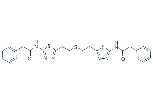
BPTES Chemical Structure
CAS: 314045-39-1
Selleck's BPTES has been cited by 52 publications
Purity & Quality Control
Batch:
Purity: >97%
97
Related compound libraries
Choose Selective Glutaminase Inhibitors
Cell Data
| Cell Lines | Assay Type | Concentration | Incubation Time | Formulation | Activity Description | PMID |
|---|---|---|---|---|---|---|
| MDA-MB-231 cell | Cytotoxicity assay | 6 days | Cytotoxicity against human MDA-MB-231 cells measured on 6th day by hemocytometry, IC50=2.61 μM | 26988803 | ||
| MDA-MB-231 | Growth inhibition assay | 72 hrs | Growth inhibition of human MDA-MB-231 cells after 72 hrs by MTS assay, IC50 = 6.8 μM. | 28609101 | ||
| Aspc-1 | Growth inhibition assay | 72 hrs | Growth inhibition of human Aspc-1 cells after 72 hrs by MTS assay, IC50 = 10.2 μM. | 28609101 | ||
| HCC827 | Cytotoxicity assay | 48 hrs | Cytotoxicity against human erlotinib-resistant HCC827 cells assessed as growth inhibition after 48 hrs by CCK8 assay, IC50 = 42.4 μM. | 28174105 | ||
| HT1080 | Cytotoxicity assay | 48 hrs | Cytotoxicity against human HT1080 cells assessed as growth inhibition after 48 hrs by CCK8 assay, IC50 = 47.72 μM. | 28174105 | ||
| Click to View More Cell Line Experimental Data | ||||||
Biological Activity
| Description | BPTES is a potent and selective Glutaminase GLS1 (KGA) inhibitor with IC50 of 0.16 μM. It has no effect on glutamate dehydrogenase activity and causes only a very slight inhibition of γ-glutamyl transpeptidase activity. | ||
|---|---|---|---|
| Targets |
|
| In vitro | ||||
| In vitro | BPTES inhibits glutaminase activity expressed in human kidney cells with IC50 of 0.18 μM, and inhibits glutamate efflux by microglia with IC50 of 80-120 nM. [1] BPTES preferentially slows cell growth in D54 cells with mutant IDH1. BPTES also inhibits glutaminase activity, lowers glutamate and α-KG levels, and increases glycolytic intermediates. [2] BPTES (10 μM) inhibits cell growth of mHCC 3–4 cells derived from LAP/MYC tumors. BPTES also inhibits growth of a MYC-dependent P493 cells by blocking DNA replication, leading to cell death and fragmentation. [3] | |||
|---|---|---|---|---|
| Kinase Assay | Glutaminase Inhibition: Cell Free Assay | |||
| Assay plates are prepared containing 2 μL test compound in DMSO/well. The enzyme is diluted to 1 unit (liver) or 0.8 unit (kidney)/100 μL in glutaminase assay buffer, and 100 μL diluted enzyme is added to each well of the assay plate by Multidrop. The contents are mixed by shaking at full speed for 1 min on TiterMix 100. The plates are preincubated at room temperature (RT) for 20 min to allow binding of test compounds to glutaminase, and 50 μL glutamine solution (7 mM in assay buffer) is added to each well by Multidrop. The contents are shaken at full speed for 30 sec on TiterMix 100, and the plates are then incubated at RT for 60 min (liver) or 90 min (kidney). To stop the reactions, 20 μL HCl (0.3 N) is added to each well by Multidrop and mixed immediately by shaking for 30 sec on TiterMix 100. For quantification, glutamate (formed by glutaminase-catalyzed hydrolysis of glutamine) is oxidized to 2-oxoglutarate by a second enzyme, glutamate dehydrogenase (GDH), with the concomitant production of the reduced form of nicotinamide adenine dinucleotide (NADH). Reduction of nitro blue tetrazolium (NBT) in the assay solution by NADH, catalyzed by phenazine methosulphate (PMS), results in the formation of a blue-purple formazan. The absorption of formazan at 540 nm is linearly proportional to the concentration of glutamate up to 200 μM. NBT/GDH reagent (50 μL) is added to each well by Multidrop and mixed by shaking for 30 sec on TiterMix 100, and the plates are incubated at RT for 20 min to allow color formation by the GDH reaction. Glutamate concentration is determined from formazan concentration as determined by reading OD540 nm on a SpectraMax 340. | ||||
| Cell Research | Cell lines | D54 cells | ||
| Concentrations | -- | |||
| Incubation Time | 48 h | |||
| Method | Cell growth assays are carried out using alamarBlue. Cells are plated at a density of 500 cells/well in a 96-well black clear bottom plate. At 24 hrs, media is changed to the appropriate media (DMEM with 4.5 g/L, 1.5 g/L or 0.1 g/L glucose, 10% FBS, pencillin/streptomycin, and 4 mM glutamine with or without doxycyline. 48 hours after plating, compounds or DMSO are added. Media and alamarBlue is added to a volume of 200 礚 in each well. Fluorescence is measured at 48 hrs (AOA, BPTES) using a Victor3 plate-reader. |
|||
| Experimental Result Images | Methods | Biomarkers | Images | PMID |
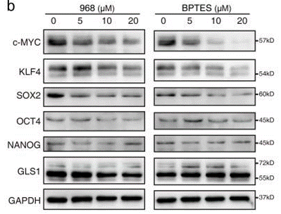
| Western blot | c-Myc / KLF4 / SOX2 / OCT4 / NANOG / GLS1 γH2AX |

|
30555042 | |
| In Vivo | ||
| In vivo | In LAP/MYC mice, BPTES (12.5 mg/kg, i.p.) prolongs survival with no significant effects on MYC, GLS, or GLS2 levels. BPTES (200 μg/mouse, i.p.) also inhibits tumor cell growth in mice harboring P493 tumor xenografts. [3] | |
|---|---|---|
| Animal Research | Animal Models | LAP/MYC mice |
| Dosages | 12.5 mg/kg | |
| Administration | i.p. | |
Chemical lnformation & Solubility
| Molecular Weight | 524.68 | Formula | C24H24N6O2S3 |
| CAS No. | 314045-39-1 | SDF | Download BPTES SDF |
| Smiles | C1=CC=C(C=C1)CC(=O)NC2=NN=C(S2)CCSCCC3=NN=C(S3)NC(=O)CC4=CC=CC=C4 | ||
| Storage (From the date of receipt) | |||
|
In vitro |
DMSO : 50 mg/mL ( (95.29 mM); Moisture-absorbing DMSO reduces solubility. Please use fresh DMSO.) Water : Insoluble Ethanol : Insoluble |
Molecular Weight Calculator |
|
In vivo Add solvents to the product individually and in order. |
In vivo Formulation Calculator |
||||
Preparing Stock Solutions
Molarity Calculator
In vivo Formulation Calculator (Clear solution)
Step 1: Enter information below (Recommended: An additional animal making an allowance for loss during the experiment)
mg/kg
g
μL
Step 2: Enter the in vivo formulation (This is only the calculator, not formulation. Please contact us first if there is no in vivo formulation at the solubility Section.)
% DMSO
%
% Tween 80
% ddH2O
%DMSO
%
Calculation results:
Working concentration: mg/ml;
Method for preparing DMSO master liquid: mg drug pre-dissolved in μL DMSO ( Master liquid concentration mg/mL, Please contact us first if the concentration exceeds the DMSO solubility of the batch of drug. )
Method for preparing in vivo formulation: Take μL DMSO master liquid, next addμL PEG300, mix and clarify, next addμL Tween 80, mix and clarify, next add μL ddH2O, mix and clarify.
Method for preparing in vivo formulation: Take μL DMSO master liquid, next add μL Corn oil, mix and clarify.
Note: 1. Please make sure the liquid is clear before adding the next solvent.
2. Be sure to add the solvent(s) in order. You must ensure that the solution obtained, in the previous addition, is a clear solution before proceeding to add the next solvent. Physical methods such
as vortex, ultrasound or hot water bath can be used to aid dissolving.
Tech Support
Answers to questions you may have can be found in the inhibitor handling instructions. Topics include how to prepare stock solutions, how to store inhibitors, and issues that need special attention for cell-based assays and animal experiments.
Tel: +1-832-582-8158 Ext:3
If you have any other enquiries, please leave a message.
* Indicates a Required Field
Tags: buy BPTES | BPTES supplier | purchase BPTES | BPTES cost | BPTES manufacturer | order BPTES | BPTES distributor


