- Bioactive Compounds
- By Signaling Pathways
- PI3K/Akt/mTOR
- Epigenetics
- Methylation
- Immunology & Inflammation
- Protein Tyrosine Kinase
- Angiogenesis
- Apoptosis
- Autophagy
- ER stress & UPR
- JAK/STAT
- MAPK
- Cytoskeletal Signaling
- Cell Cycle
- TGF-beta/Smad
- Compound Libraries
- Popular Compound Libraries
- Customize Library
- Clinical and FDA-approved Related
- Bioactive Compound Libraries
- Inhibitor Related
- Natural Product Related
- Metabolism Related
- Cell Death Related
- By Signaling Pathway
- By Disease
- Anti-infection and Antiviral Related
- Neuronal and Immunology Related
- Fragment and Covalent Related
- FDA-approved Drug Library
- FDA-approved & Passed Phase I Drug Library
- Preclinical/Clinical Compound Library
- Bioactive Compound Library-I
- Bioactive Compound Library-Ⅱ
- Kinase Inhibitor Library
- Express-Pick Library
- Natural Product Library
- Human Endogenous Metabolite Compound Library
- Alkaloid Compound LibraryNew
- Angiogenesis Related compound Library
- Anti-Aging Compound Library
- Anti-alzheimer Disease Compound Library
- Antibiotics compound Library
- Anti-cancer Compound Library
- Anti-cancer Compound Library-Ⅱ
- Anti-cancer Metabolism Compound Library
- Anti-Cardiovascular Disease Compound Library
- Anti-diabetic Compound Library
- Anti-infection Compound Library
- Antioxidant Compound Library
- Anti-parasitic Compound Library
- Antiviral Compound Library
- Apoptosis Compound Library
- Autophagy Compound Library
- Calcium Channel Blocker LibraryNew
- Cambridge Cancer Compound Library
- Carbohydrate Metabolism Compound LibraryNew
- Cell Cycle compound library
- CNS-Penetrant Compound Library
- Covalent Inhibitor Library
- Cytokine Inhibitor LibraryNew
- Cytoskeletal Signaling Pathway Compound Library
- DNA Damage/DNA Repair compound Library
- Drug-like Compound Library
- Endoplasmic Reticulum Stress Compound Library
- Epigenetics Compound Library
- Exosome Secretion Related Compound LibraryNew
- FDA-approved Anticancer Drug LibraryNew
- Ferroptosis Compound Library
- Flavonoid Compound Library
- Fragment Library
- Glutamine Metabolism Compound Library
- Glycolysis Compound Library
- GPCR Compound Library
- Gut Microbial Metabolite Library
- HIF-1 Signaling Pathway Compound Library
- Highly Selective Inhibitor Library
- Histone modification compound library
- HTS Library for Drug Discovery
- Human Hormone Related Compound LibraryNew
- Human Transcription Factor Compound LibraryNew
- Immunology/Inflammation Compound Library
- Inhibitor Library
- Ion Channel Ligand Library
- JAK/STAT compound library
- Lipid Metabolism Compound LibraryNew
- Macrocyclic Compound Library
- MAPK Inhibitor Library
- Medicine Food Homology Compound Library
- Metabolism Compound Library
- Methylation Compound Library
- Mouse Metabolite Compound LibraryNew
- Natural Organic Compound Library
- Neuronal Signaling Compound Library
- NF-κB Signaling Compound Library
- Nucleoside Analogue Library
- Obesity Compound Library
- Oxidative Stress Compound LibraryNew
- Plant Extract Library
- Phenotypic Screening Library
- PI3K/Akt Inhibitor Library
- Protease Inhibitor Library
- Protein-protein Interaction Inhibitor Library
- Pyroptosis Compound Library
- Small Molecule Immuno-Oncology Compound Library
- Mitochondria-Targeted Compound LibraryNew
- Stem Cell Differentiation Compound LibraryNew
- Stem Cell Signaling Compound Library
- Natural Phenol Compound LibraryNew
- Natural Terpenoid Compound LibraryNew
- TGF-beta/Smad compound library
- Traditional Chinese Medicine Library
- Tyrosine Kinase Inhibitor Library
- Ubiquitination Compound Library
-
Cherry Picking
You can personalize your library with chemicals from within Selleck's inventory. Build the right library for your research endeavors by choosing from compounds in all of our available libraries.
Please contact us at info@selleckchem.com to customize your library.
You could select:
- Antibodies
- Bioreagents
- qPCR
- 2x SYBR Green qPCR Master Mix
- 2x SYBR Green qPCR Master Mix(Low ROX)
- 2x SYBR Green qPCR Master Mix(High ROX)
- Protein Assay
- Protein A/G Magnetic Beads for IP
- Anti-DYKDDDDK Tag magnetic beads
- Anti-DYKDDDDK Tag Affinity Gel
- Anti-Myc magnetic beads
- Anti-HA magnetic beads
- Poly DYKDDDDK Tag Peptide lyophilized powder
- Protease Inhibitor Cocktail
- Protease Inhibitor Cocktail (EDTA-Free, 100X in DMSO)
- Phosphatase Inhibitor Cocktail (2 Tubes, 100X)
- Cell Biology
- Cell Counting Kit-8 (CCK-8)
- Animal Experiment
- Mouse Direct PCR Kit (For Genotyping)
- New Products
- Contact Us
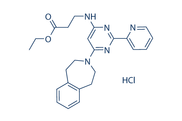
GSK J4 HCl
GSK J4 HCl is a cell permeable prodrug of GSK J1, which is the first selective inhibitor of the H3K27 histone demethylase JMJD3 and UTX with IC50 of 60 nM in a cell-free assay and inactive against a panel of demethylases of the JMJ family.

GSK J4 HCl Chemical Structure
CAS: 1797983-09-5
Selleck's GSK J4 HCl has been cited by 54 publications
Purity & Quality Control
Batch:
Purity:
99.48%
99.48
Related compound libraries
Choose Selective Histone Demethylase Inhibitors
Cell Data
| Cell Lines | Assay Type | Concentration | Incubation Time | Formulation | Activity Description | PMID |
|---|---|---|---|---|---|---|
| CUTLL1 | Growth inhibitory assay | 2 μM | DMSO | affects cell growth | 25132549 | |
| CUTLL1 | Apoptosis assay | 2 μM | DMSO | induces apoptosis | 25132549 | |
| CUTLL1 | Function assay | 2 μM | DMSO | induces cell cycle arrest | 25132549 | |
| CUTLL1 | Kinase assay | 6 μM | DMSO | leads to increased H3K27me3 | 25132549 | |
| SF7761 | Kinase assay | 6 μM | DMSO | increases K27 methylation | 25401693 | |
| SF8628 | Kinase assay | 6 μM | DMSO | increases K28 methylation | 25401693 | |
| H3.3 | Kinase assay | 6 μM | DMSO | increases K29 methylation | 25401693 | |
| SF9012 | Kinase assay | 6 μM | DMSO | increases K30 methylation | 25401693 | |
| SF9402 | Kinase assay | 6 μM | DMSO | increases K31 methylation | 25401693 | |
| SF9427 | Kinase assay | 6 μM | DMSO | increases K32 methylation | 25401693 | |
| human astrocytes | Kinase assay | 6 μM | DMSO | increases K33 methylation | 25401693 | |
| SF7761 | Growth inhibitory assay | 6 μM | DMSO | inhibits K27M glioma cell growth | 25401693 | |
| SF8628 | Growth inhibitory assay | 6 μM | DMSO | inhibits K28M glioma cell growth | 25401693 | |
| H3.3 | Growth inhibitory assay | 6 μM | DMSO | inhibits K29M glioma cell growth | 25401693 | |
| SF9012 | Growth inhibitory assay | 6 μM | DMSO | inhibits K30M glioma cell growth | 25401693 | |
| SF9402 | Growth inhibitory assay | 6 μM | DMSO | inhibits K31M glioma cell growth | 25401693 | |
| SF9427 | Growth inhibitory assay | 6 μM | DMSO | inhibits K32M glioma cell growth | 25401693 | |
| human astrocytes | Growth inhibitory assay | 6 μM | DMSO | inhibits K33M glioma cell growth | 25401693 | |
| TG neurons | Function assay | 50 μM | DMSO | inhibits HSV-1 reactivation from sensory neurons | 25552720 | |
| Th17 | Function assay | 80 nM | DMSO | inhibits cell differentiation | 25840993 | |
| β-cells | Function assay | 20 μM | DMSO | blunts IFNγ, Il-1β, and TNFα-induced chemokine gene expression | 26505193 | |
| β-cells | Function assay | 20 μM | DMSO | induces β-cell dysfunction | 26505193 | |
| ESCs | Function assay | 1.8 µM | DMSO | induces DNA damage along with activation of the DNA damage response | 26759175 | |
| Raw 264.7 | Function assay | 0.8192 µM | DMSO | inhibits TNF-α production | 26776360 | |
| MCF7 | Function assay | 1 to 10 uM | 30 hrs | Inhibition of KDM5A in human MCF7 cells assessed as effect on H3K4me3 methylation levels at 1 to 10 uM after 30 hrs by Western blot analysis | 30392349 | |
| MCF7 | Function assay | 1 to 10 uM | 30 hrs | Inhibition of KDM5A in human MCF7 cells assessed as effect on H3K27me3 methylation levels at 1 to 10 uM after 30 hrs by Western blot analysis | 30392349 | |
| RAW264.7 | 0.82 uM | 24 hrs | Inhibition of LPS-induced TNFalpha production in mouse RAW264.7 cells 0.82 uM after 24 hrs by ELISA | 26776360 | ||
| RAW264.7 | 24 hrs | Inhibition of LPS-induced TNFalpha production in mouse RAW264.7 cells after 24 hrs by ELISA | 26776360 | |||
| Click to View More Cell Line Experimental Data | ||||||
Biological Activity
| Description | GSK J4 HCl is a cell permeable prodrug of GSK J1, which is the first selective inhibitor of the H3K27 histone demethylase JMJD3 and UTX with IC50 of 60 nM in a cell-free assay and inactive against a panel of demethylases of the JMJ family. | ||
|---|---|---|---|
| Targets |
|
| In vitro | ||||
| In vitro | GSK J4 HCl is an ethyl ester derivative of the JMJD3 selective histone demethylase inhibitor GSK-J1 with an IC50 value greater than 50 μM in vitro. GSK J4 HCl is used to probe the consequences of demethylation of H3K27me3. In human primary macrophages, GSK-J4 inhibits the lipopolysaccharide-induced production of cytokines, including pro-inflammatory tumour necrosis factor (TNF). In addition, GSK-J4 prevents the lipopolysaccharide-induced loss of H3K27me3 associated with the TNF transcription start sites and blocked the recruitment of RNA polymerase II. [1] |
|||
|---|---|---|---|---|
| Kinase Assay | Histone Demethylase AlphaScreen | |||
| Inhibition of histone demethylases is assessed using the histone demethylase AlphaScreen assay (Amplified Luminescence Proximity Homogenous Assay). This assay uses a biotinylated peptide substrate and relies on detection of the product methyl mark using a specific antibody coupled to protein-A acceptor beads and a Steptavidin donor bead to capture the peptide. In brief, recombinant demethylase enzymes are incubated in the presence of Fe2+ in the form of Ferrous Ammonium Sulphate (FAS), -ketoglutarate (KG) and biotinylated peptide substrate. L-Ascorbic Acid is included to provide a reducing environment and prevent oxidation of Fe2+. After incubation with peptide substrate the presence of the product is detected using AlphaScreen technology. The demethylase AlphaScreen assays are performed in 384-well plate format using white proxiplates. All steps are carried out in assay buffer (50 mM HEPES pH 7.5, 0.1% (w/v) BSA and 0.01 % (v/v) Tween-20). FAS is dissolved fresh each day in 20 mM HCl to a concentration of 400 mM and diluted to 1.0 mM in deionized water. All other components are dissolved fresh each day in deionized water. For IC50 determinations 5 μL of assay buffer containing demethylase enzyme is transferred to wells of a 384-well proxiplate. Titrations of compound (0.1 μL) are transferred to each well and the enzymes allowed to pre-incubate for 15 minutes with compound (final concentration of DMSO is 1%). The enzyme reaction is initiated by addition of 5 μL of a substrate mix consisting of α-KG, FAS, L-Ascorbic Acid and biotinylated peptide substrate and the reaction incubated for the indicated time at room temperature. The enzyme reaction is stopped after the indicated time by addinton of 5 μL of EDTA (7.5 mM final concentration in assay buffer). Streptavidin Donor beads (0.08 mg/ml) and Protein-A conjugated acceptor beads (0.08 mg/ml) are pre-incubated for 1 hour with an antibody to the product methyl mark and the presence of biotin-H3-product is detected by addition of 5 μL of the preincubated AlphaScreen beads (final concentrations of 0.02 mg/ml with respect to acceptor and donor beads). Detection is allowed to proceed for 1 hour at room temperature and the assay plates read in a BMG Labtech Pherastar FS plate reader. Data are normalized to the no enzyme control and the IC50 determined from the nonlinear regression curve fit using GraphPad Prism 5. | ||||
| Cell Research | Cell lines | Mouse podocytes | ||
| Concentrations | 5 μM | |||
| Incubation Time | 48 h | |||
| Method | Cells were serum starved for 4 hours, followed by treatment with EPZ-6438 (10 μM) or GSK-J4 (5 μM) for 48 hours. |
|||
| In Vivo | ||
| In vivo |
GSK-J4 hydrochloride is a potent dual inhibitor of H3K27me3/me2-demethylases JMJD3/KDM6B and UTX/KDM6A. It inhibits LPS-induced TNF-α production in human primary macrophages. GSK-J4 hydrochloride is a cell permeable prodrug of GSK-J1. |
|
|---|---|---|
| Animal Research | Animal Models | BALB/c mice |
| Dosages | 10 mg/kg | |
| Administration | i.p. | |
Chemical lnformation & Solubility
| Molecular Weight | 453.96 | Formula | C24H27N5O2.HCl |
| CAS No. | 1797983-09-5 | SDF | Download GSK J4 HCl SDF |
| Smiles | CCOC(=O)CCNC1=CC(=NC(=N1)C2=CC=CC=N2)N3CCC4=CC=CC=C4CC3.Cl | ||
| Storage (From the date of receipt) | |||
|
In vitro |
DMSO : 91 mg/mL ( (200.45 mM); Moisture-absorbing DMSO reduces solubility. Please use fresh DMSO.) Ethanol : 91 mg/mL Water : 10 mg/mL |
Molecular Weight Calculator |
|
In vivo Add solvents to the product individually and in order. |
In vivo Formulation Calculator |
||||
Preparing Stock Solutions
Molarity Calculator
In vivo Formulation Calculator (Clear solution)
Step 1: Enter information below (Recommended: An additional animal making an allowance for loss during the experiment)
mg/kg
g
μL
Step 2: Enter the in vivo formulation (This is only the calculator, not formulation. Please contact us first if there is no in vivo formulation at the solubility Section.)
% DMSO
%
% Tween 80
% ddH2O
%DMSO
%
Calculation results:
Working concentration: mg/ml;
Method for preparing DMSO master liquid: mg drug pre-dissolved in μL DMSO ( Master liquid concentration mg/mL, Please contact us first if the concentration exceeds the DMSO solubility of the batch of drug. )
Method for preparing in vivo formulation: Take μL DMSO master liquid, next addμL PEG300, mix and clarify, next addμL Tween 80, mix and clarify, next add μL ddH2O, mix and clarify.
Method for preparing in vivo formulation: Take μL DMSO master liquid, next add μL Corn oil, mix and clarify.
Note: 1. Please make sure the liquid is clear before adding the next solvent.
2. Be sure to add the solvent(s) in order. You must ensure that the solution obtained, in the previous addition, is a clear solution before proceeding to add the next solvent. Physical methods such
as vortex, ultrasound or hot water bath can be used to aid dissolving.
Tech Support
Answers to questions you may have can be found in the inhibitor handling instructions. Topics include how to prepare stock solutions, how to store inhibitors, and issues that need special attention for cell-based assays and animal experiments.
Tel: +1-832-582-8158 Ext:3
If you have any other enquiries, please leave a message.
* Indicates a Required Field
Tags: buy GSK J4 HCl | GSK J4 HCl supplier | purchase GSK J4 HCl | GSK J4 HCl cost | GSK J4 HCl manufacturer | order GSK J4 HCl | GSK J4 HCl distributor


