- Bioactive Compounds
- By Signaling Pathways
- PI3K/Akt/mTOR
- Epigenetics
- Methylation
- Immunology & Inflammation
- Protein Tyrosine Kinase
- Angiogenesis
- Apoptosis
- Autophagy
- ER stress & UPR
- JAK/STAT
- MAPK
- Cytoskeletal Signaling
- Cell Cycle
- TGF-beta/Smad
- Compound Libraries
- Popular Compound Libraries
- Customize Library
- Clinical and FDA-approved Related
- Bioactive Compound Libraries
- Inhibitor Related
- Natural Product Related
- Metabolism Related
- Cell Death Related
- By Signaling Pathway
- By Disease
- Anti-infection and Antiviral Related
- Neuronal and Immunology Related
- Fragment and Covalent Related
- FDA-approved Drug Library
- FDA-approved & Passed Phase I Drug Library
- Preclinical/Clinical Compound Library
- Bioactive Compound Library-I
- Bioactive Compound Library-Ⅱ
- Kinase Inhibitor Library
- Express-Pick Library
- Natural Product Library
- Human Endogenous Metabolite Compound Library
- Alkaloid Compound LibraryNew
- Angiogenesis Related compound Library
- Anti-Aging Compound Library
- Anti-alzheimer Disease Compound Library
- Antibiotics compound Library
- Anti-cancer Compound Library
- Anti-cancer Compound Library-Ⅱ
- Anti-cancer Metabolism Compound Library
- Anti-Cardiovascular Disease Compound Library
- Anti-diabetic Compound Library
- Anti-infection Compound Library
- Antioxidant Compound Library
- Anti-parasitic Compound Library
- Antiviral Compound Library
- Apoptosis Compound Library
- Autophagy Compound Library
- Calcium Channel Blocker LibraryNew
- Cambridge Cancer Compound Library
- Carbohydrate Metabolism Compound LibraryNew
- Cell Cycle compound library
- CNS-Penetrant Compound Library
- Covalent Inhibitor Library
- Cytokine Inhibitor LibraryNew
- Cytoskeletal Signaling Pathway Compound Library
- DNA Damage/DNA Repair compound Library
- Drug-like Compound Library
- Endoplasmic Reticulum Stress Compound Library
- Epigenetics Compound Library
- Exosome Secretion Related Compound LibraryNew
- FDA-approved Anticancer Drug LibraryNew
- Ferroptosis Compound Library
- Flavonoid Compound Library
- Fragment Library
- Glutamine Metabolism Compound Library
- Glycolysis Compound Library
- GPCR Compound Library
- Gut Microbial Metabolite Library
- HIF-1 Signaling Pathway Compound Library
- Highly Selective Inhibitor Library
- Histone modification compound library
- HTS Library for Drug Discovery
- Human Hormone Related Compound LibraryNew
- Human Transcription Factor Compound LibraryNew
- Immunology/Inflammation Compound Library
- Inhibitor Library
- Ion Channel Ligand Library
- JAK/STAT compound library
- Lipid Metabolism Compound LibraryNew
- Macrocyclic Compound Library
- MAPK Inhibitor Library
- Medicine Food Homology Compound Library
- Metabolism Compound Library
- Methylation Compound Library
- Mouse Metabolite Compound LibraryNew
- Natural Organic Compound Library
- Neuronal Signaling Compound Library
- NF-κB Signaling Compound Library
- Nucleoside Analogue Library
- Obesity Compound Library
- Oxidative Stress Compound LibraryNew
- Plant Extract Library
- Phenotypic Screening Library
- PI3K/Akt Inhibitor Library
- Protease Inhibitor Library
- Protein-protein Interaction Inhibitor Library
- Pyroptosis Compound Library
- Small Molecule Immuno-Oncology Compound Library
- Mitochondria-Targeted Compound LibraryNew
- Stem Cell Differentiation Compound LibraryNew
- Stem Cell Signaling Compound Library
- Natural Phenol Compound LibraryNew
- Natural Terpenoid Compound LibraryNew
- TGF-beta/Smad compound library
- Traditional Chinese Medicine Library
- Tyrosine Kinase Inhibitor Library
- Ubiquitination Compound Library
-
Cherry Picking
You can personalize your library with chemicals from within Selleck's inventory. Build the right library for your research endeavors by choosing from compounds in all of our available libraries.
Please contact us at [email protected] to customize your library.
You could select:
- Antibodies
- Bioreagents
- qPCR
- 2x SYBR Green qPCR Master Mix
- 2x SYBR Green qPCR Master Mix(Low ROX)
- 2x SYBR Green qPCR Master Mix(High ROX)
- Protein Assay
- Protein A/G Magnetic Beads for IP
- Anti-DYKDDDDK Tag magnetic beads
- Anti-DYKDDDDK Tag Affinity Gel
- Anti-Myc magnetic beads
- Anti-HA magnetic beads
- Poly DYKDDDDK Tag Peptide lyophilized powder
- Protease Inhibitor Cocktail
- Protease Inhibitor Cocktail (EDTA-Free, 100X in DMSO)
- Phosphatase Inhibitor Cocktail (2 Tubes, 100X)
- Cell Biology
- Cell Counting Kit-8 (CCK-8)
- Animal Experiment
- Mouse Direct PCR Kit (For Genotyping)
- New Products
- Contact Us
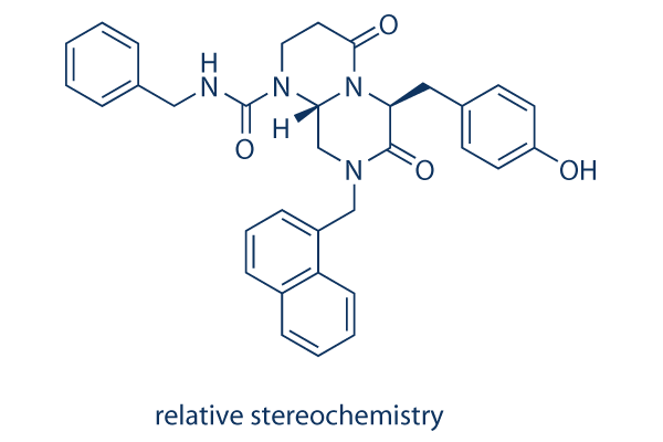
ICG-001
ICG-001 antagonizes Wnt/β-catenin/TCF-mediated transcription and specifically binds to CREB-binding protein (CBP) with IC50 of 3 μM, but is not the related transcriptional coactivator p300. ICG-001 induces apoptosis.

ICG-001 Chemical Structure
CAS: 780757-88-2 (relative stereochemistry); 847591-62-2 (absolute stereochemistry)
Selleck's ICG-001 has been cited by 182 publications
Purity & Quality Control
Batch:
Purity:
99.75%
99.75
Products often used together with ICG-001
Foscenvivint and Auranofin synergistically inhibit the growth of colon cancer in subcutaneous xenograft mice models and restrain metastasis in lung metastasis mice models.
Foscenvivint ameliorates Paclitaxel-induced increase in FOXM1 expression and cancer stem cells (CSC) phenotype as well as tumor initiation capacity in vitro.
Foscenvivint and JQ1 combination treatment induces strong cytotoxic effects in H3.3K27M-mutated diffuse intrinsic pontine gliomas (DIPG) cell lines.
Foscenvivint and Pyrvinium plus Bortezomib decrease cell viability and increase cell apoptosis rate in RPMI-8226BR and KMS-11BR cell lines.
Foscenvivint and Wnt-C59 inhibit in vitro growth of human cholangiocarcinoma cells and reduce tumor area and number in xenograft and thioacetamide models.
Related compound libraries
Choose Selective Epigenetic Reader Domain Inhibitors
Cell Data
| Cell Lines | Assay Type | Concentration | Incubation Time | Formulation | Activity Description | PMID |
|---|---|---|---|---|---|---|
| SH-SY5Y | Apoptosis Assay | 50 μm | 24 h | DMSO | blocks the protective effect of melatonin against PrP (106–126)-induced apoptotic signals | 25251028 |
| AsPC-1 | Growth Inhibition Assay | 1-20 μM | 2/4/6 d | inhibits the cell growth in a dose-dependent manner | 25082960 | |
| MiaPaCa-2 | Growth Inhibition Assay | 1-20 μM | 2/4/6 d | inhibits the cell growth in a dose-dependent manner | 25082960 | |
| PANC-1 | Growth Inhibition Assay | 1-20 μM | 2/4/6 d | inhibits the cell growth in a dose-dependent manner | 25082960 | |
| L3.6pl | Growth Inhibition Assay | 1-20 μM | 2/4/6 d | inhibits the cell growth in a dose-dependent manner | 25082960 | |
| SH-SY5Y | Apoptosis Assay | 10 μM | 24 h | inhibits the neuroprotective effects of hypoxia against PrP (106-126)-mediated neuronal cell death | 23900566 | |
| HKC-8 | Function Assay | 10 µM | 24 h | abolishes β-catenin–mediated RAS induction | 25012166 | |
| HK-2 | Function Assay | 10 µM | 3 h | reduced the expression of TGF-β1, α-SMA, and CTGF after treatment with HHE | 23690997 | |
| HepT1 | Apoptosis Assay | 0-100 μM | 24 h | IC50=34 μM | 23266718 | |
| HuH6 | Apoptosis Assay | 0-100 μM | 24 h | IC50=39 μM | 23266718 | |
| MCF7 | Function Assay | 5 μm | inhibits leptin-mediated increased expression of Snail, Slug, and Zeb2 | 22270359 | ||
| RLE-6TN | Function Assay | 2.5/5/7.5 μM | 48 h | inhibits TGF-β1-induced α-SMA induction and EMT | 22241478 | |
| HKC-8 | Function Assay | 5/10/20 μM | 48 h | blocks β-catenin-driven gene expression | 21816937 | |
| SW480 | Growth Inhibition Assay | 2-100 μM | IC50=5.8±0.68 μM | 15782138 | ||
| SW480 | Function assay | Inhibition of CBP binding to beta-casein in human SW480 cells by immunoblot analysis, IC50 = 1.3 μM. | 23232060 | |||
| A549 | Antiproliferative assay | 72 hrs | Antiproliferative activity against human A549 cells after 72 hrs by MTT assay, GI50 = 6.1 μM. | 24950489 | ||
| HepG2 | Antiproliferative assay | 72 hrs | Antiproliferative activity against human HepG2 cells after 72 hrs by MTT assay, GI50 = 12.7 μM. | 24950489 | ||
| LoVo | Antiproliferative assay | 72 hrs | Antiproliferative activity against human LoVo cells after 72 hrs by MTT assay, GI50 = 15.6 μM. | 24950489 | ||
| HT-29 | Antiproliferative assay | 72 hrs | Antiproliferative activity against human HT-29 cells after 72 hrs by MTT assay, GI50 = 17.2 μM. | 24950489 | ||
| HT29 | Function assay | 24 hrs | Inhibition of Wnt signaling in human HT29 cells assessed as inhibition of beta-catenin-mediated Tcf/Lef transcriptional activity after 24 hrs by dual luciferase reporter gene assay relative to control, IC50 = 18.7 μM. | 24950489 | ||
| TC32 | qHTS assay | qHTS of pediatric cancer cell lines to identify multiple opportunities for drug repurposing: Primary screen for TC32 cells | 29435139 | |||
| A673 | qHTS assay | qHTS of pediatric cancer cell lines to identify multiple opportunities for drug repurposing: Primary screen for A673 cells | 29435139 | |||
| DAOY | qHTS assay | qHTS of pediatric cancer cell lines to identify multiple opportunities for drug repurposing: Primary screen for DAOY cells | 29435139 | |||
| BT-37 | qHTS assay | qHTS of pediatric cancer cell lines to identify multiple opportunities for drug repurposing: Primary screen for BT-37 cells | 29435139 | |||
| RD | qHTS assay | qHTS of pediatric cancer cell lines to identify multiple opportunities for drug repurposing: Primary screen for RD cells | 29435139 | |||
| MG 63 (6-TG R) | qHTS assay | qHTS of pediatric cancer cell lines to identify multiple opportunities for drug repurposing: Primary screen for MG 63 (6-TG R) cells | 29435139 | |||
| NB1643 | qHTS assay | qHTS of pediatric cancer cell lines to identify multiple opportunities for drug repurposing: Primary screen for NB1643 cells | 29435139 | |||
| OHS-50 | qHTS assay | qHTS of pediatric cancer cell lines to identify multiple opportunities for drug repurposing: Primary screen for OHS-50 cells | 29435139 | |||
| SJ-GBM2 | qHTS assay | qHTS of pediatric cancer cell lines to identify multiple opportunities for drug repurposing: Primary screen for SJ-GBM2 cells | 29435139 | |||
| SK-N-MC | qHTS assay | qHTS of pediatric cancer cell lines to identify multiple opportunities for drug repurposing: Primary screen for SK-N-MC cells | 29435139 | |||
| NB-EBc1 | qHTS assay | qHTS of pediatric cancer cell lines to identify multiple opportunities for drug repurposing: Primary screen for NB-EBc1 cells | 29435139 | |||
| LAN-5 | qHTS assay | qHTS of pediatric cancer cell lines to identify multiple opportunities for drug repurposing: Primary screen for LAN-5 cells | 29435139 | |||
| LoVo | Cytotoxicity assay | 10 uM | 72 hrs | Cytotoxicity against Wnt/beta-catenin signalling dependent human LoVo cells assessed as cell viability at 10 uM after 72 hrs by ATPlite assay | ChEMBL | |
| NCI-H1703 | Function assay | 10 uM | 24 hrs | Inhibition of TNIK in human NCI-H1703 cells transfected with lentiviral vector 7TFP assessed as reduction of GSK3 inhibitor X activated TNIK-mediated Wnt/TCF/beta-catenin-dependent transcription at 10 uM after 24 hrs by luciferase reporter assay | ChEMBL | |
| HCT116 | Cytotoxicity assay | 10 uM | 72 hrs | Cytotoxicity against Wnt/beta-catenin signalling dependent human HCT116 cells assessed as cell viability at 10 uM after 72 hrs by ATPlite assay | ChEMBL | |
| Click to View More Cell Line Experimental Data | ||||||
Biological Activity
| Description | ICG-001 antagonizes Wnt/β-catenin/TCF-mediated transcription and specifically binds to CREB-binding protein (CBP) with IC50 of 3 μM, but is not the related transcriptional coactivator p300. ICG-001 induces apoptosis. | ||
|---|---|---|---|
| Targets |
|
| In vitro | ||||
| In vitro | ICG-001 has no effect on the related reporter construct, FOPFLASH, which contains mutated TCF sites. After treatment with 25μM of ICG-001 for 8 hours, SW480 cell reduces the steady-state levels of Survivin and Cyclin D1 RNA and protein, both of which can be up-regulated by β-catenin. ICG-001 selectively induces apoptosis in transformed cells but not in normal colon cells, reduces in vitro growth of colon carcinoma cells. [1] ICG-001, can phenotypically rescue normal nerve growth factor (NGF) -induced neuronal differentiation and neurite outgrowth in the presenilin-1 mutant cells, emphasizing the importance of the TCF/β-catenin signaling pathway on neurite outgrowth and neuronal differentiation. [2] A recent study demonstrates that 5μM ICG-001 inhibits leptin-induced EMT, invasion and tumorsphere formation in MCF7 cells. [3] |
|||
|---|---|---|---|---|
| Kinase Assay | DUAL-Luciferase Reporter Assay | |||
| The Dual-Luciferase Reporter (DLR) Assay System provides an efficient means of performing dual reporter assays. In the DLRTM Assay, the activities of firefly (Photinus pyralis) and Renilla (Renilla reniformis, also known as sea pansy) luciferases are measured sequentially from a single sample. The firefly luciferase reporter is measured first by adding Luciferase Assay Reagent II (LAR II) to generate a “glow-type” luminescent signal. After quantifying the firefly luminescence, this reaction is quenched, and the Renilla luciferase reaction is initiated by simultaneously adding Stop & Glo® Reagent to the same tube. The Stop & Glo® Reagent also produces a “glow-type” signal from the Renilla luciferase, which decays slowly over the course of the measurement. In the DLRTM Assay System, both reporters yield linear assays with subattomole (<10-18) sensitivities and no endogenous activity of either reporter in the experimental host cells. Furthermore, the integrated format of the DLRTM Assay provides rapid quantitation of both reporters either in transfected cells or in cell-free transcription/translation reactions. | ||||
| Cell Research | Cell lines | Human colon carcinoma cell lines SW480, SW620, and HCT116, normal colonic epithelial cell line CCD-841Co | ||
| Concentrations | ~25 μM | |||
| Incubation Time | 24 hours | |||
| Method | 1. Prior to starting the assay, prepare the Apo-ONE Caspase-3/7 Reagent, and mix thoroughly. 2. For best results, empirical determination of the optimal cell number, apoptosis induction treatment and incubation period for the cell culture system may be necessary. 3. Use identical cell numbers and volumes for the assay and the negative control samples. 4. Do not mix Apo-ONE Caspase-3/7 Reagent and samples by manual pipetting. Mixing in this manner is unnecessary and may create bubbles that interfere with fluorescence readings or cross-contaminate the samples. Gentle mixing may be performed using a plate shaker. 5. Total incubation time for the assay depends upon the amount of caspase- 3/7 present in the sample. 6. The Apo-ONE Caspase-3/7 Reagent is formulated to mediate cellular lysis and support optimal caspase-3/7 activity. In rare instances, the reagent does not affect complete lysis of cultured cells. In such cases, lysis is enhanced by a freeze-thaw cycle. For best results, freeze at -70 °C, then thaw at room temperature. After equilibration, mix to homogeneity and incubate until measurable fluorescence is achieved |
|||
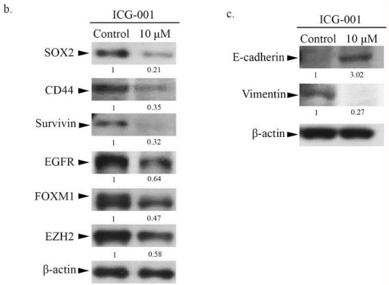
| Experimental Result Images | Methods | Biomarkers | Images | PMID |
| Western blot | SOX-2 / CD44 / Survivin / EGFR / FOXM1 / EZH2 / Vimentin pβ-catenin beta-catenin / F-actin ABC / Beta-catenin / E-cadherin / N-cadherin / MMP-9 / c-MYC / Actin / Histone H3 CCNB1 / Cyclin D1 |

|
25897700 | |
| In Vivo | ||
| In vivo |
Administration of a water-soluble analog of ICG-001 for 9 weeks reduces the formation of colon and small intestinal polyps by 42% as effectively as the nonsteroidal antiinflammatory agent Sulindac, which has consistently demonstrated efficacy in this model. No overt toxicity is detected throughout the course of treatment. In the SW620 nude mouse xenograft model of tumor regression, 150 mg/kg, i.v. of analog demonstrates a dramatic reduction in tumor volume over the 19-day course of treatment, with no mortality or weight loss. [1] ICG-001 (5 mg/kg per day) significantly inhibits beta-catenin signaling and attenuates bleomycin-induced lung fibrosis in mice, while concurrently preserving the epithelium. [4] |
|
|---|---|---|
| Animal Research | Animal Models | C57BL/6 and nude mice tumor models |
| Dosages | 20 mg/ kg | |
| Administration | i.p. | |
Chemical lnformation & Solubility
| Molecular Weight | 548.63 | Formula | C33H32N4O4 |
| CAS No. | 780757-88-2 (relative stereochemistry); 847591-62-2 (absolute stereochemistry) | SDF | Download ICG-001 SDF |
| Smiles | C1CN(C2CN(C(=O)C(N2C1=O)CC3=CC=C(C=C3)O)CC4=CC=CC5=CC=CC=C54)C(=O)NCC6=CC=CC=C6 | ||
| Storage (From the date of receipt) | |||
|
In vitro |
DMSO : 30 mg/mL ( (54.68 mM); Moisture-absorbing DMSO reduces solubility. Please use fresh DMSO.) Water : Insoluble Ethanol : Insoluble |
Molecular Weight Calculator |
|
In vivo Add solvents to the product individually and in order. |
In vivo Formulation Calculator |
||||
Preparing Stock Solutions
Molarity Calculator
In vivo Formulation Calculator (Clear solution)
Step 1: Enter information below (Recommended: An additional animal making an allowance for loss during the experiment)
mg/kg
g
μL
Step 2: Enter the in vivo formulation (This is only the calculator, not formulation. Please contact us first if there is no in vivo formulation at the solubility Section.)
% DMSO
%
% Tween 80
% ddH2O
%DMSO
%
Calculation results:
Working concentration: mg/ml;
Method for preparing DMSO master liquid: mg drug pre-dissolved in μL DMSO ( Master liquid concentration mg/mL, Please contact us first if the concentration exceeds the DMSO solubility of the batch of drug. )
Method for preparing in vivo formulation: Take μL DMSO master liquid, next addμL PEG300, mix and clarify, next addμL Tween 80, mix and clarify, next add μL ddH2O, mix and clarify.
Method for preparing in vivo formulation: Take μL DMSO master liquid, next add μL Corn oil, mix and clarify.
Note: 1. Please make sure the liquid is clear before adding the next solvent.
2. Be sure to add the solvent(s) in order. You must ensure that the solution obtained, in the previous addition, is a clear solution before proceeding to add the next solvent. Physical methods such
as vortex, ultrasound or hot water bath can be used to aid dissolving.
Tech Support
Answers to questions you may have can be found in the inhibitor handling instructions. Topics include how to prepare stock solutions, how to store inhibitors, and issues that need special attention for cell-based assays and animal experiments.
Tel: +1-832-582-8158 Ext:3
If you have any other enquiries, please leave a message.
* Indicates a Required Field
Frequently Asked Questions
Question 1:
If the compound is stored in DMSO at -80, how long would it be stable? For cell culture, how long should I change for the fresh medium with ICG-001?
Answer:
The product in DMSO solution can be stored at 4 degree for 1 week and -20 degree for 1 month. The best storage condition is solid powder, even at -80 the solution is not stable enough for long term storage. For cell culture, you need change medium every 48h.
Tags: buy ICG-001 | ICG-001 supplier | purchase ICG-001 | ICG-001 cost | ICG-001 manufacturer | order ICG-001 | ICG-001 distributor


