- Bioactive Compounds
- By Signaling Pathways
- PI3K/Akt/mTOR
- Epigenetics
- Methylation
- Immunology & Inflammation
- Protein Tyrosine Kinase
- Angiogenesis
- Apoptosis
- Autophagy
- ER stress & UPR
- JAK/STAT
- MAPK
- Cytoskeletal Signaling
- Cell Cycle
- TGF-beta/Smad
- Compound Libraries
- Popular Compound Libraries
- Customize Library
- Clinical and FDA-approved Related
- Bioactive Compound Libraries
- Inhibitor Related
- Natural Product Related
- Metabolism Related
- Cell Death Related
- By Signaling Pathway
- By Disease
- Anti-infection and Antiviral Related
- Neuronal and Immunology Related
- Fragment and Covalent Related
- FDA-approved Drug Library
- FDA-approved & Passed Phase I Drug Library
- Preclinical/Clinical Compound Library
- Bioactive Compound Library-I
- Bioactive Compound Library-Ⅱ
- Kinase Inhibitor Library
- Express-Pick Library
- Natural Product Library
- Human Endogenous Metabolite Compound Library
- Alkaloid Compound LibraryNew
- Angiogenesis Related compound Library
- Anti-Aging Compound Library
- Anti-alzheimer Disease Compound Library
- Antibiotics compound Library
- Anti-cancer Compound Library
- Anti-cancer Compound Library-Ⅱ
- Anti-cancer Metabolism Compound Library
- Anti-Cardiovascular Disease Compound Library
- Anti-diabetic Compound Library
- Anti-infection Compound Library
- Antioxidant Compound Library
- Anti-parasitic Compound Library
- Antiviral Compound Library
- Apoptosis Compound Library
- Autophagy Compound Library
- Calcium Channel Blocker LibraryNew
- Cambridge Cancer Compound Library
- Carbohydrate Metabolism Compound LibraryNew
- Cell Cycle compound library
- CNS-Penetrant Compound Library
- Covalent Inhibitor Library
- Cytokine Inhibitor LibraryNew
- Cytoskeletal Signaling Pathway Compound Library
- DNA Damage/DNA Repair compound Library
- Drug-like Compound Library
- Endoplasmic Reticulum Stress Compound Library
- Epigenetics Compound Library
- Exosome Secretion Related Compound LibraryNew
- FDA-approved Anticancer Drug LibraryNew
- Ferroptosis Compound Library
- Flavonoid Compound Library
- Fragment Library
- Glutamine Metabolism Compound Library
- Glycolysis Compound Library
- GPCR Compound Library
- Gut Microbial Metabolite Library
- HIF-1 Signaling Pathway Compound Library
- Highly Selective Inhibitor Library
- Histone modification compound library
- HTS Library for Drug Discovery
- Human Hormone Related Compound LibraryNew
- Human Transcription Factor Compound LibraryNew
- Immunology/Inflammation Compound Library
- Inhibitor Library
- Ion Channel Ligand Library
- JAK/STAT compound library
- Lipid Metabolism Compound LibraryNew
- Macrocyclic Compound Library
- MAPK Inhibitor Library
- Medicine Food Homology Compound Library
- Metabolism Compound Library
- Methylation Compound Library
- Mouse Metabolite Compound LibraryNew
- Natural Organic Compound Library
- Neuronal Signaling Compound Library
- NF-κB Signaling Compound Library
- Nucleoside Analogue Library
- Obesity Compound Library
- Oxidative Stress Compound LibraryNew
- Plant Extract Library
- Phenotypic Screening Library
- PI3K/Akt Inhibitor Library
- Protease Inhibitor Library
- Protein-protein Interaction Inhibitor Library
- Pyroptosis Compound Library
- Small Molecule Immuno-Oncology Compound Library
- Mitochondria-Targeted Compound LibraryNew
- Stem Cell Differentiation Compound LibraryNew
- Stem Cell Signaling Compound Library
- Natural Phenol Compound LibraryNew
- Natural Terpenoid Compound LibraryNew
- TGF-beta/Smad compound library
- Traditional Chinese Medicine Library
- Tyrosine Kinase Inhibitor Library
- Ubiquitination Compound Library
-
Cherry Picking
You can personalize your library with chemicals from within Selleck's inventory. Build the right library for your research endeavors by choosing from compounds in all of our available libraries.
Please contact us at [email protected] to customize your library.
You could select:
- Antibodies
- Bioreagents
- qPCR
- 2x SYBR Green qPCR Master Mix
- 2x SYBR Green qPCR Master Mix(Low ROX)
- 2x SYBR Green qPCR Master Mix(High ROX)
- Protein Assay
- Protein A/G Magnetic Beads for IP
- Anti-DYKDDDDK Tag magnetic beads
- Anti-DYKDDDDK Tag Affinity Gel
- Anti-Myc magnetic beads
- Anti-HA magnetic beads
- Poly DYKDDDDK Tag Peptide lyophilized powder
- Protease Inhibitor Cocktail
- Protease Inhibitor Cocktail (EDTA-Free, 100X in DMSO)
- Phosphatase Inhibitor Cocktail (2 Tubes, 100X)
- Cell Biology
- Cell Counting Kit-8 (CCK-8)
- Animal Experiment
- Mouse Direct PCR Kit (For Genotyping)
- New Products
- Contact Us
RITA
Synonyms: NSC 652287
RITA induces both DNA-protein and DNA-DNA cross-links with no detectable DNA single-strand breaks, and also inhibits MDM2-p53 interaction by targeting p53.

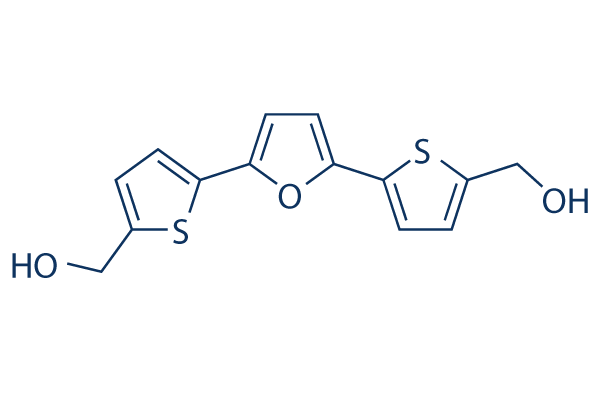
RITA Chemical Structure
CAS: 213261-59-7
Selleck's RITA has been cited by 11 Publications
1 Customer Review
Purity & Quality Control
Other p53 Products
Related compound libraries
Choose Selective p53 Inhibitors
Biological Activity
| Description | RITA induces both DNA-protein and DNA-DNA cross-links with no detectable DNA single-strand breaks, and also inhibits MDM2-p53 interaction by targeting p53. | ||
|---|---|---|---|
| Features | Inducer of DNA cross-links, not a DNA intercalator. | ||
| Targets |
|
| In vitro | ||||
| In vitro | RITA shows a highly selective pattern of differential cytotoxic activity in the tumor cell lines, due to cellular accumulation to the cytosolic (S100) fraction. RITA also inhibits the growth of other renal cell lines including ACHN and UO-31 with IC50 of 13 μM and 37 μM, respectively. [1] RITA (10 nM) causes cell cycle arrest with accumulation of cells at the G2-M phase and induces DNA fragmentation and apoptosis at 100 nM, both with evaluated p53 protein levels. RITA (30 nM) also induces both DNA-protein and DNA-DNA cross-links in A498 cells. Meanwhile RITA has no effects on top1-mediated relaxation of supercoiled SV40 DNA. [2] RITA significantly suppresses the growth of HCT116 cells (97%) but only slightly inhibits the growth of HCT116 TP53-/- cells (13%). RITA is much more efficient at growth suppression in wild-type p53-expressing tumor cell lines than in cell lines lacking p53 and those expressing mutant p53. RITA binds full-length p53 but not glutathione S-transferase (GST) protein or HDM-2 (a key regulator of p53 is strongly supported by the rescue of embryonic lethality of MDM2). RITA blocks p53−HDM-2 interaction and p53 ubiquitination. RITA substantially decreases the amount of HDM-2 that is co-precipitated with p53, although both proteins are upregulated. RITA prevents interactions between the purified GST-p53 and 6XHis-tagged His-HDM-2 proteins. [3] RITA is shown to induce apoptosis by promoting p53Ser46 phosphorylation. [4] RITA induces activation of p53 in conjunction with up-regulation of phosphorylated ASK-1, MKK-4 and c-Jun. RITA induces the activation of JNK signaling. [5] But On the contrary, another results by nuclear magnetic resonance (NMR) show that RITA does not block the formation of the complex between p53 (residues 1-312) and the N-terminal p53-binding domain of MDM2 (residues 1-118), which is highly probable that the binding of RITA requires native conformation of p53. [6] | |||
|---|---|---|---|---|
| Cell Research | Cell lines | HCT116 cells and HCT116 TP53-/- cells | ||
| Concentrations | 0.1 nM - 1 mM, 10 mM stocked in DMSO | |||
| Incubation Time | 48 hours | |||
| Method | Examination to assess susceptibility of cells to RITA (0.1 nM - 1 mM) is done using the XTT assay. Cells are inoculated into 96-well flat-bottom plates at a density of 1500 cells per well and incubated for 24 hours at 37 °C in a humidified 5% CO2 5% air atmosphere. Serial concentrations of RITA in DMSO are added to the wells, and sensitivity is determined 48 hours after the addition of RIT | |||
| In Vivo | ||
| In vivo | RITA is well tolerated in mice after intraperitoneal administration, with no observable weight loss at doses up to 10 mg/kg during 1 month. After five injections of 0.1 mg/kg of RITA, the growth of the HCT116 tumors is suppressed by 40%, without apparent effects on the HCT116 TP53-/- tumors. At a dose of 1 or 10 mg/kg, RITA shows strong antitumor activity. Five 1 mg/kg injections of RITA results in a more than twofold decrease in the growth rate of p53-positive xenografts without any effect on p53-null xenografts. HCT116 tumors are 90% smaller in mice treated with 10 mg/kg of RITA than in control untreated mice. RITA inhibits the tumor growth in a wild-type p53−dependent manner. [3] | |
|---|---|---|
| Animal Research | Animal Models | SCID mice carrying HCT116 and HCT116 TP53-/- xenografts |
| Dosages | 0.1 mg/kg, 1 mg/kg or 10 mg/kg | |
| Administration | Administered via i.v. or i.p. | |
| NCT Number | Recruitment | Conditions | Sponsor/Collaborators | Start Date | Phases |
|---|---|---|---|---|---|
| NCT05260203 | Completed | Multiple Myeloma|Solitary Plasmacytoma|Amyloidosis|Chronic Myeloid Leukemia|Chronic Lymphocytic Leukemia|T Cell Non-Hodgkin Lymphoma|Lymphocytic Lymphoma|Hodgkin Lymphoma|B-cell Non Hodgkin Lymphoma|Acute Myeloid Leukemia|Myelodysplasia|Chronic Myeloproliferative Disorder|Treatment Adherence|Treatment Adherence and Compliance | Advice Pharma Group srl | June 4 2022 | Not Applicable |
| NCT05153551 | Completed | Autism Spectrum Disorder | University of Michigan|Blue Cross Blue Shield of Michigan Foundation | January 27 2022 | Not Applicable |
| NCT03806127 | Completed | Irritable Bowel Syndrome | Urovant Sciences GmbH | December 31 2018 | Phase 2 |
| NCT00779025 | Completed | Coitus | Johnson & Johnson Consumer and Personal Products Worldwide | January 2008 | Not Applicable |
Chemical lnformation & Solubility
| Molecular Weight | 292.37 | Formula | C14H12O3S2 |
| CAS No. | 213261-59-7 | SDF | Download RITA SDF |
| Smiles | C1=C(SC(=C1)C2=CC=C(O2)C3=CC=C(S3)CO)CO | ||
| Storage (From the date of receipt) | |||
|
In vitro |
DMSO : 58 mg/mL ( (198.37 mM); Moisture-absorbing DMSO reduces solubility. Please use fresh DMSO.) Ethanol : 8 mg/mL Water : Insoluble |
Molecular Weight Calculator |
|
In vivo Add solvents to the product individually and in order. |
In vivo Formulation Calculator |
||||
Preparing Stock Solutions
Molarity Calculator
In vivo Formulation Calculator (Clear solution)
Step 1: Enter information below (Recommended: An additional animal making an allowance for loss during the experiment)
mg/kg
g
μL
Step 2: Enter the in vivo formulation (This is only the calculator, not formulation. Please contact us first if there is no in vivo formulation at the solubility Section.)
% DMSO
%
% Tween 80
% ddH2O
%DMSO
%
Calculation results:
Working concentration: mg/ml;
Method for preparing DMSO master liquid: mg drug pre-dissolved in μL DMSO ( Master liquid concentration mg/mL, Please contact us first if the concentration exceeds the DMSO solubility of the batch of drug. )
Method for preparing in vivo formulation: Take μL DMSO master liquid, next addμL PEG300, mix and clarify, next addμL Tween 80, mix and clarify, next add μL ddH2O, mix and clarify.
Method for preparing in vivo formulation: Take μL DMSO master liquid, next add μL Corn oil, mix and clarify.
Note: 1. Please make sure the liquid is clear before adding the next solvent.
2. Be sure to add the solvent(s) in order. You must ensure that the solution obtained, in the previous addition, is a clear solution before proceeding to add the next solvent. Physical methods such
as vortex, ultrasound or hot water bath can be used to aid dissolving.
Tech Support
Answers to questions you may have can be found in the inhibitor handling instructions. Topics include how to prepare stock solutions, how to store inhibitors, and issues that need special attention for cell-based assays and animal experiments.
Tel: +1-832-582-8158 Ext:3
If you have any other enquiries, please leave a message.
* Indicates a Required Field
Tags: buy RITA | RITA supplier | purchase RITA | RITA cost | RITA manufacturer | order RITA | RITA distributor


