- Bioactive Compounds
- By Signaling Pathways
- PI3K/Akt/mTOR
- Epigenetics
- Methylation
- Immunology & Inflammation
- Protein Tyrosine Kinase
- Angiogenesis
- Apoptosis
- Autophagy
- ER stress & UPR
- JAK/STAT
- MAPK
- Cytoskeletal Signaling
- Cell Cycle
- TGF-beta/Smad
- Compound Libraries
- Popular Compound Libraries
- Customize Library
- Clinical and FDA-approved Related
- Bioactive Compound Libraries
- Inhibitor Related
- Natural Product Related
- Metabolism Related
- Cell Death Related
- By Signaling Pathway
- By Disease
- Anti-infection and Antiviral Related
- Neuronal and Immunology Related
- Fragment and Covalent Related
- FDA-approved Drug Library
- FDA-approved & Passed Phase I Drug Library
- Preclinical/Clinical Compound Library
- Bioactive Compound Library-I
- Bioactive Compound Library-Ⅱ
- Kinase Inhibitor Library
- Express-Pick Library
- Natural Product Library
- Human Endogenous Metabolite Compound Library
- Alkaloid Compound LibraryNew
- Angiogenesis Related compound Library
- Anti-Aging Compound Library
- Anti-alzheimer Disease Compound Library
- Antibiotics compound Library
- Anti-cancer Compound Library
- Anti-cancer Compound Library-Ⅱ
- Anti-cancer Metabolism Compound Library
- Anti-Cardiovascular Disease Compound Library
- Anti-diabetic Compound Library
- Anti-infection Compound Library
- Antioxidant Compound Library
- Anti-parasitic Compound Library
- Antiviral Compound Library
- Apoptosis Compound Library
- Autophagy Compound Library
- Calcium Channel Blocker LibraryNew
- Cambridge Cancer Compound Library
- Carbohydrate Metabolism Compound LibraryNew
- Cell Cycle compound library
- CNS-Penetrant Compound Library
- Covalent Inhibitor Library
- Cytokine Inhibitor LibraryNew
- Cytoskeletal Signaling Pathway Compound Library
- DNA Damage/DNA Repair compound Library
- Drug-like Compound Library
- Endoplasmic Reticulum Stress Compound Library
- Epigenetics Compound Library
- Exosome Secretion Related Compound LibraryNew
- FDA-approved Anticancer Drug LibraryNew
- Ferroptosis Compound Library
- Flavonoid Compound Library
- Fragment Library
- Glutamine Metabolism Compound Library
- Glycolysis Compound Library
- GPCR Compound Library
- Gut Microbial Metabolite Library
- HIF-1 Signaling Pathway Compound Library
- Highly Selective Inhibitor Library
- Histone modification compound library
- HTS Library for Drug Discovery
- Human Hormone Related Compound LibraryNew
- Human Transcription Factor Compound LibraryNew
- Immunology/Inflammation Compound Library
- Inhibitor Library
- Ion Channel Ligand Library
- JAK/STAT compound library
- Lipid Metabolism Compound LibraryNew
- Macrocyclic Compound Library
- MAPK Inhibitor Library
- Medicine Food Homology Compound Library
- Metabolism Compound Library
- Methylation Compound Library
- Mouse Metabolite Compound LibraryNew
- Natural Organic Compound Library
- Neuronal Signaling Compound Library
- NF-κB Signaling Compound Library
- Nucleoside Analogue Library
- Obesity Compound Library
- Oxidative Stress Compound LibraryNew
- Plant Extract Library
- Phenotypic Screening Library
- PI3K/Akt Inhibitor Library
- Protease Inhibitor Library
- Protein-protein Interaction Inhibitor Library
- Pyroptosis Compound Library
- Small Molecule Immuno-Oncology Compound Library
- Mitochondria-Targeted Compound LibraryNew
- Stem Cell Differentiation Compound LibraryNew
- Stem Cell Signaling Compound Library
- Natural Phenol Compound LibraryNew
- Natural Terpenoid Compound LibraryNew
- TGF-beta/Smad compound library
- Traditional Chinese Medicine Library
- Tyrosine Kinase Inhibitor Library
- Ubiquitination Compound Library
-
Cherry Picking
You can personalize your library with chemicals from within Selleck's inventory. Build the right library for your research endeavors by choosing from compounds in all of our available libraries.
Please contact us at [email protected] to customize your library.
You could select:
- Antibodies
- Bioreagents
- qPCR
- 2x SYBR Green qPCR Master Mix
- 2x SYBR Green qPCR Master Mix(Low ROX)
- 2x SYBR Green qPCR Master Mix(High ROX)
- Protein Assay
- Protein A/G Magnetic Beads for IP
- Anti-DYKDDDDK Tag magnetic beads
- Anti-DYKDDDDK Tag Affinity Gel
- Anti-Myc magnetic beads
- Anti-HA magnetic beads
- Poly DYKDDDDK Tag Peptide lyophilized powder
- Protease Inhibitor Cocktail
- Protease Inhibitor Cocktail (EDTA-Free, 100X in DMSO)
- Phosphatase Inhibitor Cocktail (2 Tubes, 100X)
- Cell Biology
- Cell Counting Kit-8 (CCK-8)
- Animal Experiment
- Mouse Direct PCR Kit (For Genotyping)
- New Products
- Contact Us
Elacridar (GF120918)
Synonyms: GW120918, GG918, GW0918
Elacridar (GF120918, GW120918, GG918, GW0918) is a potent P-gp (MDR-1) and BCRP inhibitor.

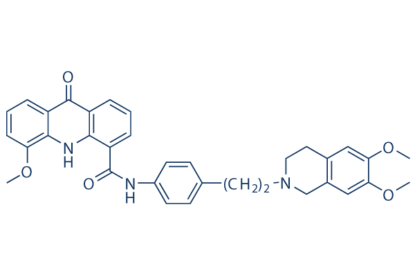
Elacridar (GF120918) Chemical Structure
CAS: 143664-11-3
Selleck's Elacridar (GF120918) has been cited by 15 Publications
3 Customer Reviews
Purity & Quality Control
Batch:
Purity:
99.97%
99.97
Products often used together with Elacridar (GF120918)
Elacridar and Imatinib induce apoptosis, reduce cell proliferation rate, and arrest CML cells in the S phase.
Elacridar and Sunitinib inhibit ABCG2 function and enhance the cytotoxic effects of sunitinib in 786-O cells.
Elacridar and Ramucirumab can restore the inhibitory action of Paclitaxel on cell cycle progression in resistant GC cells.
Elacridar and Temozolomide (TMZ) increase TMZ brain penetration and antitumor efficacy in wild-type mice.
Nanoparticles loaded with Doxorubicin and Elacridar are highly effective against HepG2 and HepG2-TS cells.
Chen D, et al. Int J Nanomedicine. 2018 Oct 25;13:6855-6870.
Other P-gp Products
Related compound libraries
Choose Selective P-gp Inhibitors
Cell Data
| Cell Lines | Assay Type | Concentration | Incubation Time | Formulation | Activity Description | PMID |
|---|---|---|---|---|---|---|
| SKVLB1 | Cytotoxicity assay | Cytotoxicity against human multidrug-resistant SKVLB1 cells in presence of adriamycin, IC50=0.02μM | 11754602 | |||
| SKVLB1 | Cytotoxicity assay | Cytotoxicity against human multidrug-resistant SKVLB1 cells, IC50=1.4μM | 11754602 | |||
| SKOV3 | Cytotoxicity assay | Cytotoxicity against human SKOV3 cells, IC50=8.1μM | 11754602 | |||
| Caco-2 | Function assay | Inhibition of Pgp measured as inhibition of [3H]vinblastine basolateral to apical transport in Caco-2 cells, EC50=2μM | 17064079 | |||
| HEK293 | Function assay | Inhibition of ABCG2 expressed in HEK293 cells assessed as mitoxantrone-mediated efflux by flow cytometry, IC50=0.41μM | 17317193 | |||
| Caco-2 | Function assay | Inhibition of human Pgp mediated [3H]vinblastine transport in human Caco-2 cells, EC50=2μM | 17936633 | |||
| Caco-2 | Function assay | Inhibition of human P-glycoprotein mediated [3H]vinblastine transport in human Caco-2 cells, EC50=2μM | 18257545 | |||
| Caco-2 | Function assay | Inhibition of human P-gp mediated [3H]vinblastine transport activity in human Caco-2 cells, EC50=2μM | 18276145 | |||
| Caco-2 | Function assay | Inhibition of ABCB1-mediated [3H]vinblastine transportation in human Caco-2 cells, IC50=2μM | 19053888 | |||
| KBv1 | Function assay | Inhibition of ABCB1 overexpressed in human KBv1 cells by flow cytometric-based calcein-AM efflux assay, IC50=0.193μM | 19170519 | |||
| MCF7/Topo | Function assay | Inhibition of ABCG2 overexpressed in human MCF7/Topo cells by flow cytometric-based mitoxantrone efflux assay, IC50=0.25μM | 19170519 | |||
| MCF7 MX | Function assay | Inhibition of BCRP expressed in MCF7 MX cells by Hoechst 33342 staining, IC50=0.4μM | 19932960 | |||
| MDCK | Function assay | Inhibition of BCRP expressed in MDCK cells by pheophorbide A assay, IC50=0.43μM | 19932960 | |||
| MDCK2-MDR1 | Function assay | 60 mins | Inhibition of human Pgp overexpressed in human MDCK2-MDR1 cells assessed as inhibition of R123 efflux after 60 mins, IC50=0.4μM | 21419632 | ||
| MCF7/Topo | Function assay | Inhibition of ABCG2 expressed in human MCF7/Topo cells by Hoechst microplate assay, IC50=0.127μM | 21570282 | |||
| Kb-V1 | Function assay | 10 mins | Inhibition of ABCB1 expressed in Kb-V1 cells after 10 mins by calcein-AM assay, IC50=0.193μM | 21570282 | ||
| Caco2 | Function assay | 3 uM | 30 mins | Inhibition of P-gp-mediated [3H]-digoxin transport in human Caco2 cells at 3 uM after 30 mins | 22266779 | |
| Caco2 | Function assay | 3 uM | 30 mins | Inhibition of BCRP-mediated [3H]estrone-3-sulfate transport in human Caco2 cells at 3 uM after 30 mins | 22266779 | |
| CCRF-CEM/VCR1000 | Function assay | Inhibition of P-glycoprotein-mediated daunorubicin efflux from human CCRF-CEM/VCR1000 cells after 240 secs by FACS flow cytometric analysis, IC50=0.07244μM | 22452412 | |||
| MCF7/Topo | Function assay | 2 hrs | Inhibition of ABCG2 in human MCF7/Topo cells after 2 hrs by Hoechst 33342 microplate assay, IC50=0.127μM | 24900683 | ||
| KBV1 | Function assay | 10 mins | Inhibition of ABCB1 in human KBV1 cells after 10 mins by Calcein-AM microplate assay, IC50=0.193μM | 24900683 | ||
| A673 | qHTS assay | qHTS of pediatric cancer cell lines to identify multiple opportunities for drug repurposing: Primary screen for A673 cells | 29435139 | |||
| SK-N-MC | qHTS assay | qHTS of pediatric cancer cell lines to identify multiple opportunities for drug repurposing: Primary screen for SK-N-MC cells | 29435139 | |||
| SK-N-SH | qHTS assay | qHTS of pediatric cancer cell lines to identify multiple opportunities for drug repurposing: Primary screen for SK-N-SH cells | 29435139 | |||
| NB1643 | qHTS assay | qHTS of pediatric cancer cell lines to identify multiple opportunities for drug repurposing: Primary screen for NB1643 cells | 29435139 | |||
| primary glioblastoma stem cells | Function assay | 10 to 10000 nM | 3 hrs | Inhibition of MDR1 in human primary glioblastoma stem cells assessed as increase in intracellular doxorubicin accumulation at 10 to 10000 nM after 3 hrs by spectrofluorimetric analysis | 30584838 | |
| primary mesothelioma stem cells | Function assay | 10 to 10000 nM | 3 hrs | Inhibition of MDR1 in human primary mesothelioma stem cells assessed as increase in intracellular doxorubicin accumulation at 10 to 10000 nM after 3 hrs by spectrofluorimetric analysis | 30584838 | |
| primary glioblastoma stem cells | Function assay | 1 uM | 72 hrs | Inhibition of MDR1 in human primary glioblastoma stem cells assessed as increase in doxorubicin-induced reduction in cell viability at 1 uM after 72 hrs by ATPlite luminescence assay | 30584838 | |
| primary mesothelioma stem cells | Function assay | 1 uM | 72 hrs | Inhibition of MDR1 in human primary mesothelioma stem cells assessed as increase in doxorubicin-induced reduction in cell viability at 1 uM after 72 hrs by ATPlite luminescence assay | 30584838 | |
| primary glioblastoma stem cells | Function assay | 1 uM | 24 hrs | Inhibition of MDR1 in human primary glioblastoma stem cells assessed as increase in doxorubicin-induced reduction in cell viability at 1 uM after 24 hrs by by LDH release assay | 30584838 | |
| primary mesothelioma stem cells | Function assay | 1 uM | 24 hrs | Inhibition of MDR1 in human primary mesothelioma stem cells assessed as increase in doxorubicin-induced reduction in cell viability at 1 uM after 24 hrs by by LDH release assay | 30584838 | |
| Click to View More Cell Line Experimental Data | ||||||
Biological Activity
| Description | Elacridar (GF120918, GW120918, GG918, GW0918) is a potent P-gp (MDR-1) and BCRP inhibitor. | ||
|---|---|---|---|
| Targets |
|
| In vitro | ||||
| In vitro | Elacridar inhibits P-glycoprotein (P-gp) labeling by [3H]azidopine with a IC50 of 0.16 μM. [2] In Caki-1 and ACHN cells, elacridar ( 2.5 μM) significantly ihibits the cell growth. The P-glycoprotein activity is found to be inhibited by elacridar. The combination of elacridar and sunitinib lead to a significant reduction in ABC Sub-family B Member 2 (ABCG2) expression in 786-O cells. [3] | |||
|---|---|---|---|---|
| Kinase Assay | Photoaffinity radiolabeling of P-gp | |||
| 10 μL of unlabeled cell membrane suspension (at 0.4 mg of protein/mL) are aliquoted into each well in 96-well plates. 5 μL of GF120918 are then added to each well. The plate is incubated 25 min at 25℃ in the dark. 5 μL of tritiated azidopine (1.8 TBq/mmol) (0.6 μM in HCI 0.2 mM) are added to each well. After 25 min of incubation at 25℃ in the dark, samples are simultaneously irradiated for 2 min at 254 nm at 0℃ with a thin layer chromatography-designed UV lamp directly in contact with the plate. Samples are solubilized in sodium dodecyl sulfate-polyacrylamide gel electrophoresis sample buffer but not heated. After separation on a 7.5% polyacrylamide gel, the gel is treated for fluorography with Amplify and exposed during 3 days onto a photosensitive film. The fluorography is analysed using a Camag thin layer chromatography Scanner II densitometer. | ||||
| Cell Research | Cell lines | ACHN, Caki-1, 786-O, and MCF-7 cells | ||
| Concentrations | ~ 5 mM | |||
| Incubation Time | 48 h | |||
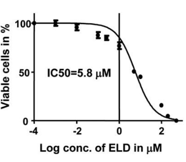
| Method | 3.0×10³ cells per well are seeded in a 96-well plate. After 24 h incubation, an optimum concentration gradient of elacridar is added to each well. After culturing for 48 h, cell viability is assessed using the proliferation reagent, MTT. Control cells are treated with the vehicle only, 0.1% DMSO. After this final incubation, the medium is aspirated and precipitated formazan crystals are dissolved in DMSO (100 μL/well). The absorbance of each well is measured at 540 nm, and a reference wavelength of 650 nm is read with a multiskan JX microplate reader. Cell viability is calculated as percentage of the control value. | |||
| Experimental Result Images | Methods | Biomarkers | Images | PMID |
| Growth inhibition assay | Cell viability |

|
25862759 | |
| In Vivo | ||
| In vivo | Oral co-administration of elacridar (100 mg/kg, p.o.) and crizotinib increases the plasma and brain concentrations and brain-to-plasma ratios of crizotinib in wild-type mice, equaling the levels in Abcb1a/1b; Abcg2-/- mice. [1] In friend leukemia virus stain B mice, the brain-to-plasm partition coefficient (Kp, brain) of elacridar is 0.82, 0.43, and 4.31 after intravenous (2.5 mg/kg), intraperitoneal (100 mg/kg), and oral (100 mg/kg) treatment, respectively. [4] In Mrp4(-/-) mice, elacridar fully inhibits P-gp mediated transport of topotecan, without siginificant effects on Bcrp1-mediated transport. [5] | |
|---|---|---|
| Animal Research | Animal Models | Male wild-type, Abcb1a/1b-/-34, Abcg2 -/-32 and Abcb1a/1b;Abcg2 |
| Dosages | 100 mg/kg | |
| Administration | p.o. | |
Chemical lnformation & Solubility
| Molecular Weight | 563.64 | Formula | C34H33N3O5 |
| CAS No. | 143664-11-3 | SDF | Download Elacridar (GF120918) SDF |
| Smiles | COC1=CC=CC2=C1NC3=C(C2=O)C=CC=C3C(=O)NC4=CC=C(C=C4)CCN5CCC6=CC(=C(C=C6C5)OC)OC | ||
| Storage (From the date of receipt) | |||
|
In vitro |
DMSO : 100 mg/mL ( (177.41 mM); Moisture-absorbing DMSO reduces solubility. Please use fresh DMSO.) Water : Insoluble Ethanol : Insoluble |
Molecular Weight Calculator |
|
In vivo Add solvents to the product individually and in order. |
In vivo Formulation Calculator |
||||
Preparing Stock Solutions
Molarity Calculator
In vivo Formulation Calculator (Clear solution)
Step 1: Enter information below (Recommended: An additional animal making an allowance for loss during the experiment)
mg/kg
g
μL
Step 2: Enter the in vivo formulation (This is only the calculator, not formulation. Please contact us first if there is no in vivo formulation at the solubility Section.)
% DMSO
%
% Tween 80
% ddH2O
%DMSO
%
Calculation results:
Working concentration: mg/ml;
Method for preparing DMSO master liquid: mg drug pre-dissolved in μL DMSO ( Master liquid concentration mg/mL, Please contact us first if the concentration exceeds the DMSO solubility of the batch of drug. )
Method for preparing in vivo formulation: Take μL DMSO master liquid, next addμL PEG300, mix and clarify, next addμL Tween 80, mix and clarify, next add μL ddH2O, mix and clarify.
Method for preparing in vivo formulation: Take μL DMSO master liquid, next add μL Corn oil, mix and clarify.
Note: 1. Please make sure the liquid is clear before adding the next solvent.
2. Be sure to add the solvent(s) in order. You must ensure that the solution obtained, in the previous addition, is a clear solution before proceeding to add the next solvent. Physical methods such
as vortex, ultrasound or hot water bath can be used to aid dissolving.
Tech Support
Answers to questions you may have can be found in the inhibitor handling instructions. Topics include how to prepare stock solutions, how to store inhibitors, and issues that need special attention for cell-based assays and animal experiments.
Tel: +1-832-582-8158 Ext:3
If you have any other enquiries, please leave a message.
* Indicates a Required Field
Tags: buy Elacridar (GF120918) | Elacridar (GF120918) supplier | purchase Elacridar (GF120918) | Elacridar (GF120918) cost | Elacridar (GF120918) manufacturer | order Elacridar (GF120918) | Elacridar (GF120918) distributor


